Japanese
English
- 有料閲覧
- Abstract 文献概要
- 1ページ目 Look Inside
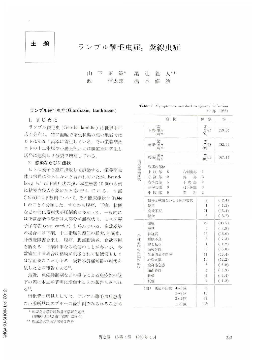
ランブル鞭毛虫症(Giardiasis,lambliasis)
1.はじめに
ランブル鞭毛虫(Giardia lamblia)は世界中に広く分布し,特に温暖で衛生状態の悪い地域ではヒトにかなり高率に寄生している.その栄養型はヒトの十二指腸や小腸上部および胆道系に寄生し活発に運動し2分裂で増殖している.
Giardiasis
1. This disease is widely distributed throughout the world and the worms live in the duodenum, upper part of the small intestine and biliary tract.
2. Symptoms are mostly related to the digestive tract; among all, abdominal pain or diarrhea.
3. X-ray picture is slight sprue pattern and nonidiopathic.
4. Diagnosis depends on the demonstration of in the diarrheal feces or the duodenal juice. This disease has become rarer, but it is still important as one of the imported helminthiasis.
Strongyloidiasis
Strongyloidiasis is most often seen in the southern part of Kyushu and Okinawa, but recently it has been reported in other districts of Japan. We have studied 42 such cases.
1. Full sensation of the abdomen and loose stool accounted for most of the clinical symptoms.
2. In the plain film of the abdomen accumulation of the air was often seen. Sometimes accumulation of the intestinal juice was seen instead.
3. Barium-filled picture showed hypertrophy or disappearance of the mucosal folds, enlargement, narrowing or stricture of the intestinal loop, and rigidity of the margins. The biliary tract was also visualized. Double contrast picture also revealed disappearance of the Kerckring's folds, irregular network shadows, granular shadows, irregular barium flecks of various size, indentations, or pseudo-diverticular deformations of the intestinal loop. They were all asymmetric.
4. These abnormal findings were most prominent in the duodenum and the upper part of the jejunum.
5. Endoscopically were recognized hypertrophy or disappearance of the duodenal mucosal folds. The mucosal surface showed white, turbid granules, edema, reddening, erosions, bleeding and ulcers.
Copyright © 1983, Igaku-Shoin Ltd. All rights reserved.