Japanese
English
- 有料閲覧
- Abstract 文献概要
- 1ページ目 Look Inside
はじめに
潰瘍性大腸炎の精密かつ周到な診断は,詳細な病歴の聴取と糞便を中心とする一般検査のほかに,X線検査・内視鏡検査・病理組織学的検査の三者の有機的な結合によりはじめて可能である1)2).
とくに内視鏡検査は,潰瘍性大腸炎の好発する直腸・下部S状結腸の検索には極めて重要な意義を有し,その診断確定には直腸・S状結腸直達鏡検査は絶対に欠かすことはできない.
一方,少数ではあるが,直腸・下部S状結腸には全く病変のみられない型の潰瘍性大腸炎もある.また,直腸S状結腸鏡で観察できる場合でも,その範囲は限局されるので,長大な大腸に波及した病態を正しく把握することは不可能である.これが深部大腸粘膜の観察が強く望まれる所以である.直腸S状結腸直達鏡による深部大腸粘膜観察の試みは,Regenbogenによりなされてはいるが3),姑息的なものであり,その観察可能の範囲もわずか50cmくらいにすぎない.
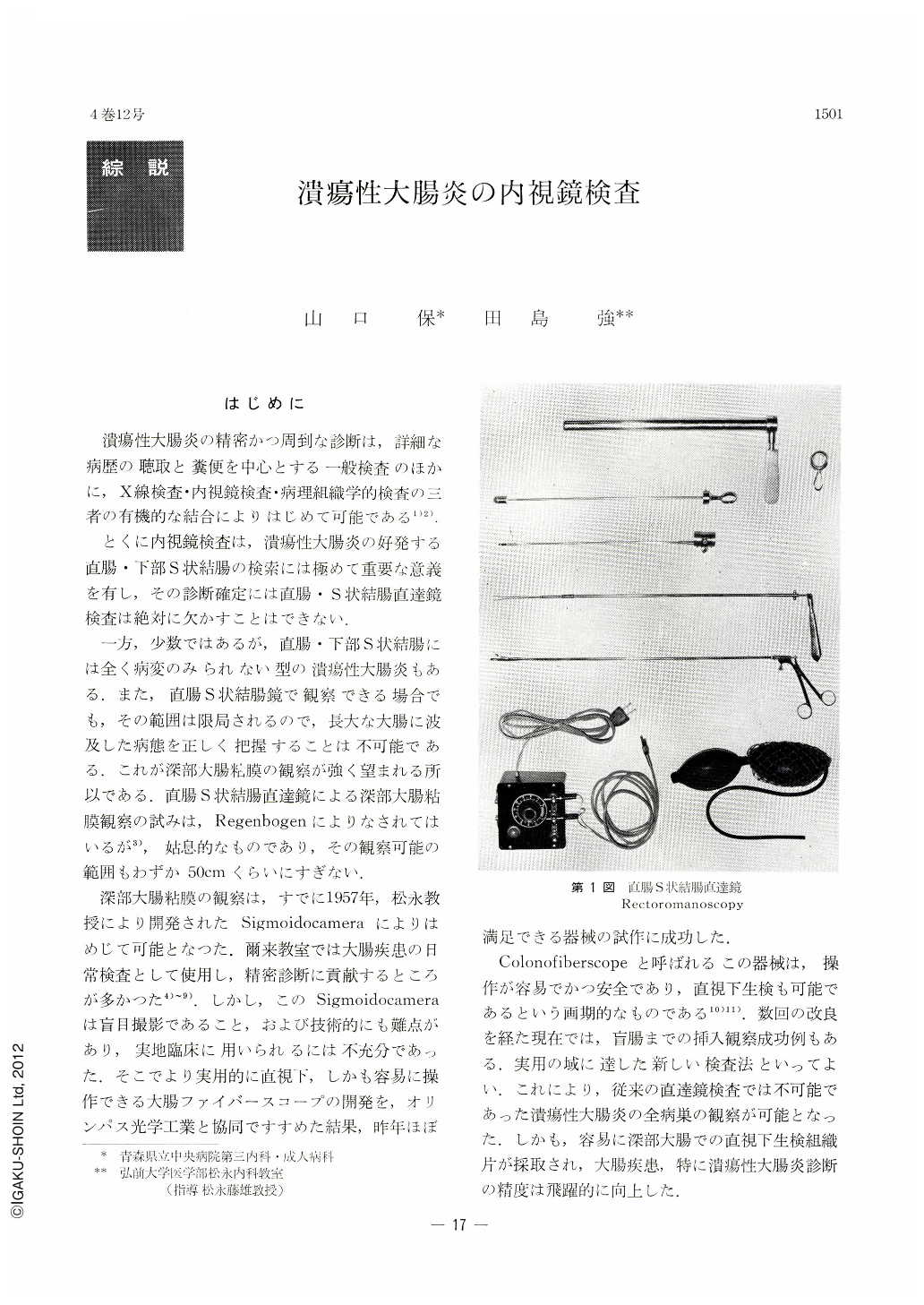
深部大腸粘膜の観察は,すでに1957年,松永教授により開発されたSigmoidocameraによりはめじて可能となった.爾来教室では大腸疾患の日常検査として使用し,精密診断に貢献するところが多かつた4)~9).しかし,このSigmoidocameraは盲目撮影であること,および技術的にも難点があり,実地臨床に用いられるには不充分であった.そこでより実用的に直視下,しかも容易に操作できる大腸ファイバースコープの開発を,オリンパス光学工業と協同ですすめた結果,昨年ほぼ満足できる器械の試作に成功した.
Colonofiberscopeと呼ばれるこの器械は,操作が容易でかつ安全であり,直視下生検も可能であるという画期的なものである10)11).数回の改良を経た現在では,盲腸までの挿入観察成功例もある.実用の域に達した新しい検査法といってよい.これにより,従来の直達鏡検査では不可能であった潰瘍性大腸炎の全病巣の観察が可能となった.しかも,容易に深部大腸での直視下生検組織片が採取され,大腸疾患,特に潰瘍性大腸炎診断の精度は飛躍的に向上した.
本稿では,直腸S状結腸直達鏡検査,Sigmoidocamera検査およびColonofiberscopyについてそれぞれの実際の手技を簡単に記し,これら内視鏡検査での潰瘍性大腸炎の所見を症例をあげて略述し,さらに鑑別診断にふれる.
It goes without saying that in the diagnosis of ulcerative colitis, x-ray examination and endoscopic study are both indispensable like two wheels of a cart; of more importance than any other diagnostic method. This is especially true in endoscopic findings as they furnish the most important materials for determination of the actual aspect and stage of this disease. However, even when exact findings can be obtained by recto-sigmoidoscopy, its extent or depth of examination is confined at most up to the flexul-a recto-sigmoidalis, being unable to photograph or observe under direct view the segment oral beyond the said flexura. Although a preliminary step in photographying the mucosa all over the colon has been initiated by the sigmoidocamera devised by Prof. Matsunaga, it involves many drawbacks such as blind photograph and difficult technique in inserting this camerra in a deeper segment. All these difficulties are now obviated in the present colonofiberscope, enabling the examiner not only to observe and photograph the mucosa of the colon under direct view, but to perform repeatedly biopsy as well, with a rise in the ratio of successful insertion of this instrument into a far deeper segment of the bowel. Of 1.85 m length, the colonofiberscope now being employed is able to reach, over beyond the sigmoid, the descending colon in 74 per cent of cases examined, whereas the sigmoid camera has only been successful in 25 per cent. In addition to this, observation and photography of the mucosa of the cecum and the ascending colon is now going to be feasible. For the diagnosis of right-side ulcerative colitis, relatively localized changes in the ascending colon and of mucosal changes of the rectum in mild cases, the colonofiberscope is a most appropriate diagnostic measure. This fact has been outlined in this paper in each type of ulcerative colitis in addition to several illustrative cases. Brief comments have also been made on practical methods of endoscopic examination of the colon, including the colonofiberscope.

Copyright © 1969, Igaku-Shoin Ltd. All rights reserved.


