Japanese
English
- 有料閲覧
- Abstract 文献概要
- 1ページ目 Look Inside
胃に原発する扁平上皮癌は極めてまれである.われわれは,30歳女性の胃体上部小彎より後壁に発生した Borrmann 2 型扁平上皮癌の1例を経験したので,報告する.
Primary pure squamous cell carcinoma of the stomach which is free from esophageal mucosa, is a rare disease. We reviewed ten cases in Japan.
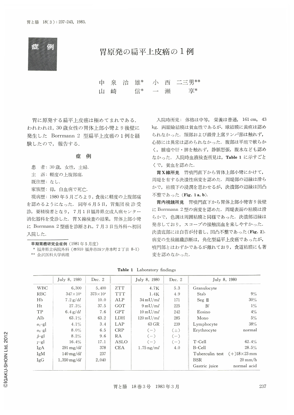
One of the patients was a 30-year-old woman. Her mother had died of leukemia. Abnormality of the stomach was pointed out when she was examined by x-ray during mass screening of the stomach because of mild stomachaches on June 5, 1980.
On July 2, she consulted our clinic and was admitted to our hospital with a diagnosis of advanced gastric cancer.
Physical examination revealed epigastric tenderness but otherwise no objective signs were found.
X-ray examination revealed a large lesion with a crater like the Borrmann 2-type cancer on the posterior wall of the upper body of the stomach.
Endoscopic examination of the stomach showed a large irregular crater with protruded margin on the posterior wall near the cardia, but esophageal mucosa was intact. Biopsy specimen from the lesion was diagnosed histologically as squamous cell carcinoma of the stomach.
On July 14, 1980, total gastrectomy, splenectomy, and Roux-Y esophagojejunostomy were performed for the patient.
The neoplasma was 6×5.5 cm in size and was on the posterior wall of the resected stomach near the cardia and the shape was macroscopically Borrmann 2-type cancer. It was 5 mm between the tumor and esophageal mucosa.
The histological diagnosis was keratinizing pure squamous cell carcinoma of the stomach-sq, INF, aw(-), ow(-), ly0, v0, n(-) (0/19)
She became healthy and left our hospital on August 9, 1980. About four months later, she was readmitted to our hospital because of general fatigue. Her laboratory data revealed hepatic dysfunction.
Her celiac angiography pointed out multiple metastasis of the liver on January 12, 1981. In spite of the fact that her liver function was marked better after two times of one-shot therapy (MMC, Bleomycin, Predonisolon) per celiac artery, she died on May 3, 1981, because of massive hematoemesis (298 days after operation).
Histogenesis of pure squamous cell carcinoma of stomach are already well discussed in various reports. However, there are no methods to unravel the exact histogenesis of the lesion.
In our case, there was a minimal lesion of the undifferentiated carcinoma in the gastric mucosa at the margin of the squamous cell carcinoma. We regarded the undifferentiated carcinoma cell as the origin of squamous cell carcinoma of the stomach.
Copyright © 1983, Igaku-Shoin Ltd. All rights reserved.