Japanese
English
- 有料閲覧
- Abstract 文献概要
- 1ページ目 Look Inside
- サイト内被引用 Cited by
胃の結節性肉芽性病変には,胃結核,胃梅毒,胃サルコイド,胃クローン病などがあげられる.このうち前二者については,凝固壊死をきたす肉芽結節であり,起炎菌の証明や血清学的検査などで鑑別される.しかしサルコイドとクローン病については,両者の鑑別は困難であり,病因についても種々の問題を残している.また胃ザルコイドは,全身サルコイド症の部分症と,金身症状を欠き,胃に限局するサノレコイドとの間にもいろいろな相違点があげられる1)~4).
われわれは,胃限局サ1レコイドの1例を経験したので報告する.
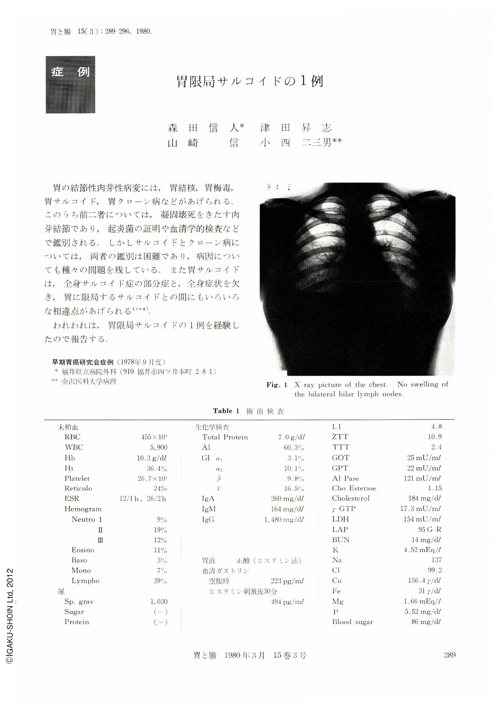
The patient. a 23 year-old nurse, with hypochromic anemia, visited our hospital for closer examination. X-ray and endoscopic examination of the stomach revealed (1) irregular giant mucosal folds on the entire wall of the corpus and antrum, (2) an open ulcer of the anterior wall at the angle, (3) and multiple disseminated verrucous lesions. Preoperative diagnosis was malignant lymphoma or diffuse carcinoma of the stomach. Total gastrectomy and splenectomy were performed with extirpation of the regional lymph nodes.
Pathologically, predominant features were thickening and granulomatous infiltration of the whole gastric wall with giant rugae and open ulcer. These granulomas consisting of epithelioid cells and Langhans type giant cells were almost uniform-sized, non-caseating, and distributed mainly in the submucosa.
Scattered granulomas were also seen in the duodenal mucosa, submucosa of the esophagus and spleen. The same numerous granulomas were also observed in all the resected lymph nodes, 59 in number.
Acid fast bacilli and fungus bodies were negative. Postoperatively tuberculin reaction was positive and kveim reaction was negative.
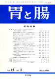
As any skin lesion, swelling of superficial lymph node and bilateral hilar lymph adenopathy were not observed in the course of this case, the present case was diagnosed as isolated gastric sarcoid.
Copyright © 1980, Igaku-Shoin Ltd. All rights reserved.