Japanese
English
- 有料閲覧
PPV購入案内
1,320円 (1,200円+税10%) こちらは、643ページから650ページの文献です。購入には医書.jp本会員登録が必要です。
会員登録完了後に改めて上記「購入サイトへ」を選択してください。
または「購入サイトへ」選択後に「購入ログイン」、「新規会員登録」の順に進んでください。
会員登録について詳しくはをご確認ください。
- Abstract 文献概要
- 1ページ目 Look Inside
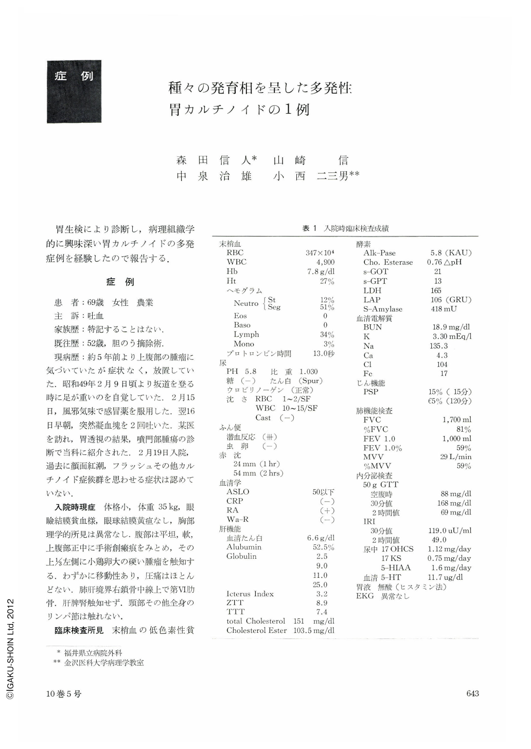
胃生検により診断し,病理組織学的に興味深い胃カルチノイドの多発症例を経験したので報告する.
A case of three independent carcinoid tumors of the stomach is described. The patient, a woman 69 years of age, had noticed since three years before a tumor in the upper part of the abdomen. It was with a chief complaint of hematemesis that she visited the hospital. Preoperative diagnosis of gastric carcinoid was established by biopsy. Prior to surgical intervention there was on increase of 5-HIAA in the urine or of 5-HT in the blood.
Subsequent to cardiectomy the excised specimen revealed three carcinoid lesions in the cardiac segment. The lesion A, measuring 3.5×2.5×2.5 cm, was localized in the submucosal layer and was lobulated. The lesion B, measuring 0.5×0.5×0.1 cm, was shaped like a dumbbell straddling the mucosal and submucosal layers. The lesion C, 0.4×0.4×0.2 cm in dimensions, was limited within the mucosal layer. Histologically three lesions were fundamentally of the same nature, showing various arrangement of cell nests that consisted of uniform cells with small almost round nuclei of very limited atypicality. We arrived at a diagnosis of carcinoid tumors of the stomach. The cells were positive for argyrophil reaction and negative for argentaffin reaction.
These three lesions each in a different stage of development seem to suggest possible patterns of histologic development of gastric carcinoid:
1. Carcinoid develops in the mucosa.
2. In early stage of development it spreads to the submucosa.
3. Most of Carcinoid tumors clinically managed show characteristics of submucosal tumor.
Effectiveness of gastric biopsy is emphasized with some reference to the literature on multiple Carcinoid tumors of the stomach.
Copyright © 1975, Igaku-Shoin Ltd. All rights reserved.