Japanese
English
- 有料閲覧
- Abstract 文献概要
- 1ページ目 Look Inside
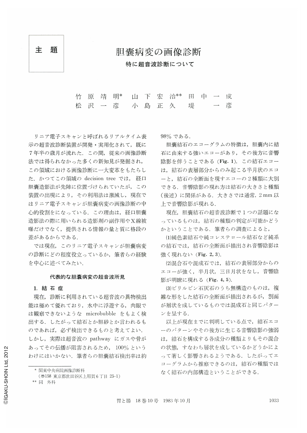
リニア電子スキャンと呼ばれるリアルタイム表示の超音波診断装置が開発・実用化されて,既に7年半の歳月が流れた.この間,従来の画像診断法では得られなかった多くの新知見が発掘され,この領域における画像診断に一大変革をもたらした.かつてこの領域のdecision treeでは,経口胆囊造影法が先陣に位置づけられていたが,この装置の出現により,その利用法は激減し,現在ではリニア電子スキャンが胆囊病変の画像診断の中心的役割をになっている.この理由は,経口胆囊造影法の際に用いられる造影剤の副作用やX線被曝だけでなく,提供される情報の量と質に格段の差があるからである.
では現在,このリニア電子スキャンが胆囊病変の診断にどの程度役立っているか,筆者らの経験を中心に述べてみたい.
Image diagnosis of gallbladder with special reference to ultrasonography was discussed.
First, echographic characteristics and differential diagnosis of gallbladder diseases including gallstone, polypoid lesion, cholecystitis and cancer of gallbladder were discussed.
Second, a role of ultrasonographic study in image diagnosis was discussed in these fields. From the experience of health check system and field system of ultrasonic mass-screening for the abdomen in Iki and Izena islands, the ultrasonography was confirmed to be absolutely indicated for the primary examination but it was also confirmed to be useful as secondary examination including further test or close examination. It was also emphasized that the indispensable test for cases which needs follow-ups.
Third, the following three points were found to be important for performing ultrasonography. Namely, scanning technique, preparation and artifact peculiar to the examination. These points are important because they influence its detecting and diagnostic abilities. Therefore, ultrasonography should be performed in good condition (early morning with fasting state) and it should be studied carefully from the two directions including r. subcostal scanning and r. intracostal scanning.
Artifacts in ultrasonogram include multiple reflection and side lobe, however these artifacts do not occur at random but occur according to a rule. The former is easy to be distinguished but the latter is difficult to do so and needs some experiences. Cultivating a professional eye is a fundamental thing for all image diagnoses.

Copyright © 1983, Igaku-Shoin Ltd. All rights reserved.


