Japanese
English
- 有料閲覧
- Abstract 文献概要
- 1ページ目 Look Inside
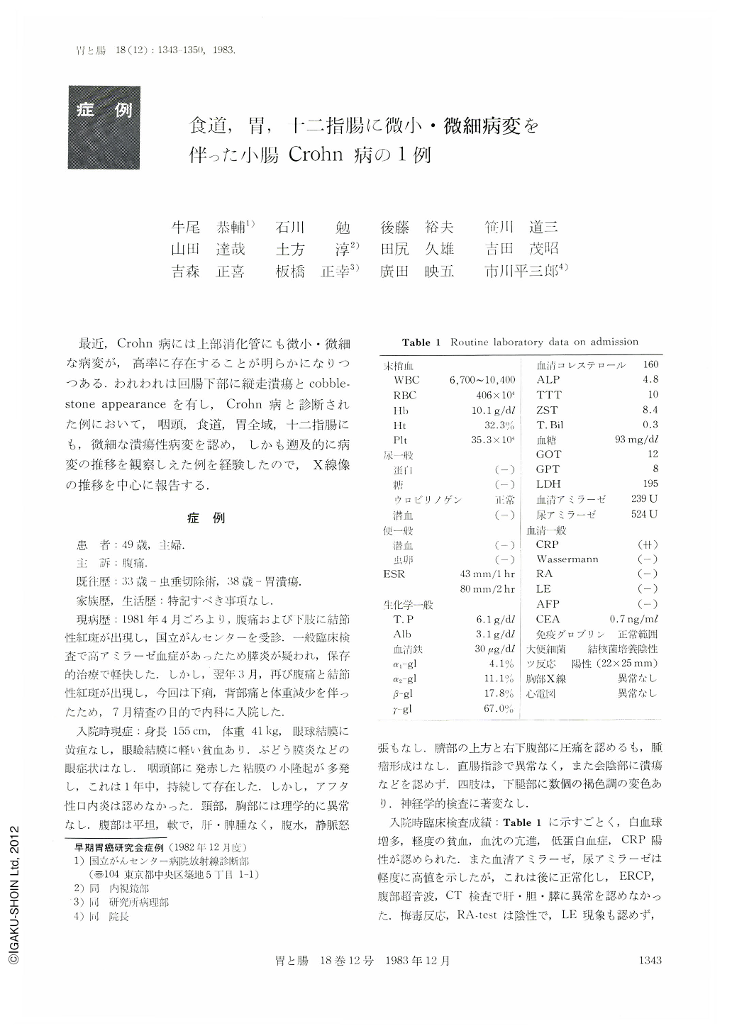
最近,Crohn病には上部消化管にも微小・微細な病変が,高率に存在することが明らかになりつつある,われわれは回腸下部に縦走潰瘍とcobblestone appearanceを有し,Crohn病と診断された例において,咽頭,食道,胃全域,十二指腸にも,微細な潰瘍性病変を認め,しかも遡及的に病変の推移を観察しえた例を経験したので,X線像の推移を中心に報告する.
症 例
患 者:49歳,主婦.
主 訴:腹痛.
既往歴:33歳-虫垂切除術,38歳-胃潰瘍.
家族歴,生活歴:特記すべき事項なし.
現病歴:1981年4月ごろより,腹痛および下肢に結節性紅斑が出現し,国立がんセンターを受診.一般臨床検査で高アミラーゼ血症があったため膵炎が疑われ,保存的治療で軽快した.しかし,翌年3月,再び腹痛と結節性紅斑が出現し,今回は下痢,背部痛と体重減少を伴ったため,7月精査の目的で内科に入院した.
Crohn's disease may involve any part of the gastrointestinal tract. However, case reports which involved the buccal mucosa, esophagus, stomach and duodenum are quite rare. We experienced one case of Crohn's disease in which minute nodular and ulcerative lesions were found in the upper gastrointestinal tract including buccal mucosa. The patient, a 49-year-old woman was admitted to our hospital complaining of abdominal pain. Barium enema and double contrast radiogram of the small intestine showed the typical findings characteristic of Crohn's disease such as longitudinal ulcer 20cm in length, cobblestone appearance and scattered aphthoid ulcers in the terminal ileum. Routine laboratory tests showed a hemoglobin of 10.1g/dl, hematocrit of 32.3%, red cell count of 406×104/mm3, positive CRP test (++), sedimentation rate 43mm/hr and hypoproteinemia (total protein 6.1g/dl, albumin 3.1g/dl). In this case, examination of double contrast radiograph and endoscopy were performed. As a result, multiple small irregular ulcerations and/or erosions with nodular mucosa were recognized in the larynx and esophagus as well as in the stomach and duodenum. Histologically, prominent inflammatory cell infiltration with accumulation of histiocyte was seen in the biopsy specimens taken from the larynx, esophagus, stomach, duodenum and ileum. Furthermore, noncaseating epithelioid cell granuloma with multinuclear giant cell was confirmed in the biopsy specimen taken from the gastric antrum.

Copyright © 1983, Igaku-Shoin Ltd. All rights reserved.


