Japanese
English
- 有料閲覧
- Abstract 文献概要
- 1ページ目 Look Inside
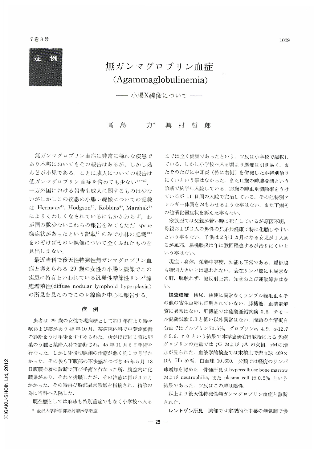
無ガンマグロブリン血症は非常に稀れな疾患であり本邦においてもその報告はあるが,しかし殆んどが小児である.ことに成人についての報告は低ガンマグロブリン血症を含めても少ない1)~5).一方外国における報告も成人に関するものは少ないがしかしこの疾患の小腸レ線像についての記載はHermans6),Hodgson7),Robbins8),Marshak9)によりくわしくなされているにもかかわらず,わが国の数少ないこれらの報告をみてもただsprue様症状があったという記載1)のみで小林の記載10)をのぞけばそのレ線像について全くふれたものを見出しえない.
最近当科で後天性特発性無ガンマグロブリン血症と考えられる29歳の女性の小腸レ線像でこの疾患に特有といわれている汎発性結節性リンパ濾胞増殖性(diffuse nodular lymphoid hyperplasia)の所見を見たのでこのレ線像を中心に報告する.
Because of infrequent recognition of agammaglobulinemia, only a few cases have been reported in Japan. Even despite the attention to the specificity of clinical and laboratory findings, little has been written concerning the roentgenologic features of the small intestine in this disease.
A 29-year-old woman had a diagnosis of acquired idiopathic agammaglobulinemia, which was based on a characteristic history and the electrophoretic pattern. On satisfactory roentgenograms of the small intestine, the mucosa appears to be studded with innumerable, tiny polypoid masses ranging from 1 to 3 mm in diameter and uniformly distributed throughout the small intestine and right colon. The filling defects are round and regular in outline.
Normal gammaglobulin levels may be found in many of those patients who have selective deficiencies of gammaglobulin. Also, susceptibility to infection has not been a feature of milder cases. So the pattern, diffuse nodular lymphoid hyperplasia of the small intestine, which can be easily recognized roentgenologically, may be an important clue to detecting this rare disease.

Copyright © 1972, Igaku-Shoin Ltd. All rights reserved.


