Japanese
English
- 有料閲覧
- Abstract 文献概要
- 1ページ目 Look Inside
- サイト内被引用 Cited by
小腸の多発性輪状狭窄は腸結核に多くみられる所見で,クローン病ではまれである.われわれは,多発性輪状狭窄を伴った小腸クローン病の1例を経験し,ほぼ全病変を小腸二重造影,術後造影,切除標本において対比しえたので報告する.
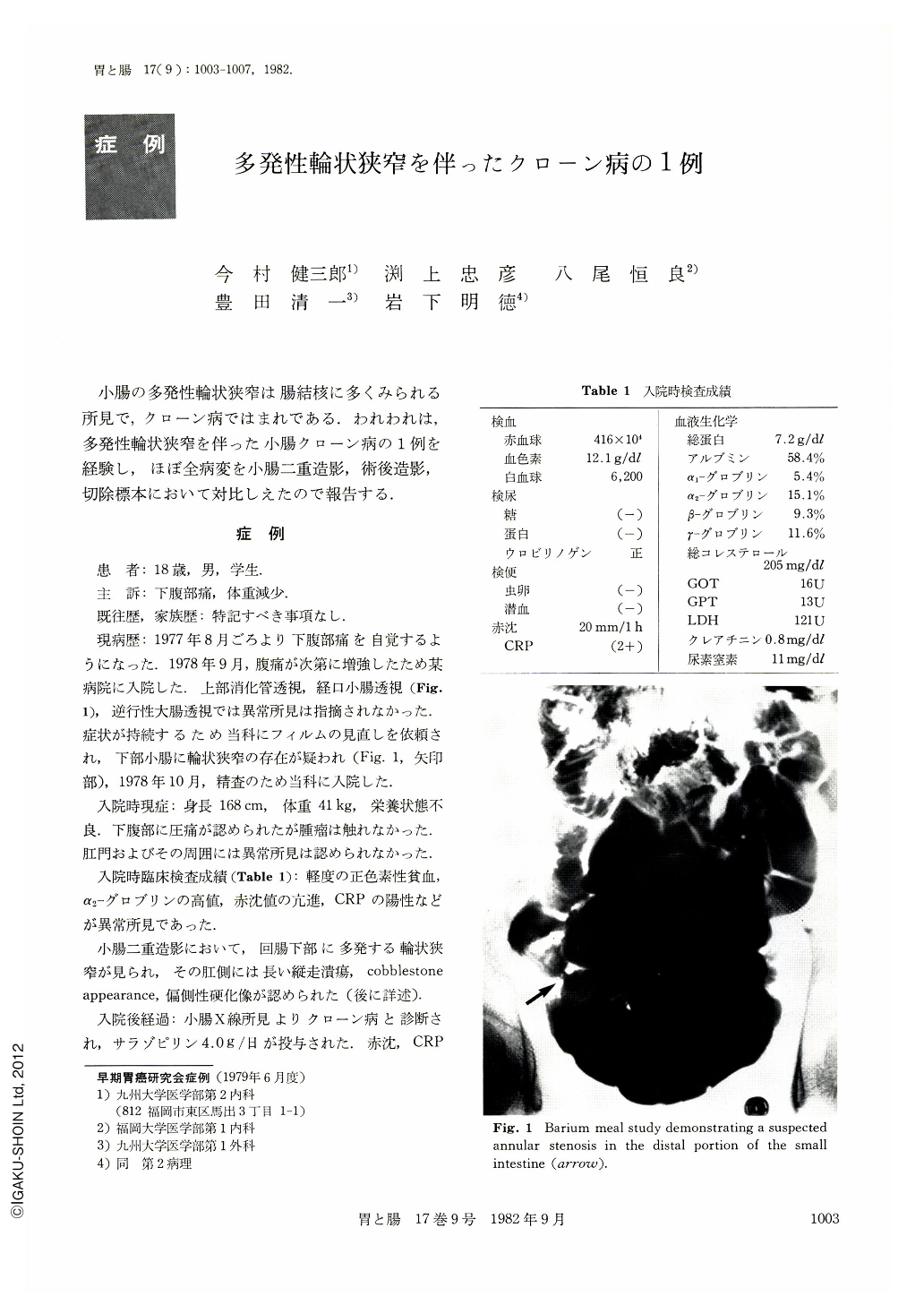
An 18-year-old man patient with Crohn's disease is reported. He was admitted to a hospital in September 1978 with complaints of lower abdominal pain and weight loss. On barium meal study of the small intestine no abnormal lesion could be pointed out, since the distal intestinal loop was conglomerated in the pelvic cavity. On review of those films by us, an annular stenosis of the distal ileum was suspected, and he was referred to us in October 1978 for further examinations. On double contrast study of the small intestine multiple annular stenoses, longitudinal ulcers, cobblestone appearance and eccentric rigidity were found. Crohn's disease was strongly suspected despite the presence of multiple annular stenoses. He underwent a resection of the diseased segment in February 1979 and the diagnosis was histopathologically proved. Comparative study among the resected specimen, postoperative roentgenograms and double contrast study were done. Most of the lesions including a shallow and small ulcer could be well judged by comparison.
Although multiple annular stenoses are regarded as characteristic of intestinal tuberculosis, the diagnosis as Crohn's disease in the presented case could only be made by the presence of eccentric rigidity, longitudinal ulcers and cobblestone appearance. It seems important to try to wholly demonstrate the intestinal loop in x-ray examination of the small intestine.

Copyright © 1982, Igaku-Shoin Ltd. All rights reserved.


