Japanese
English
- 有料閲覧
- Abstract 文献概要
- 1ページ目 Look Inside
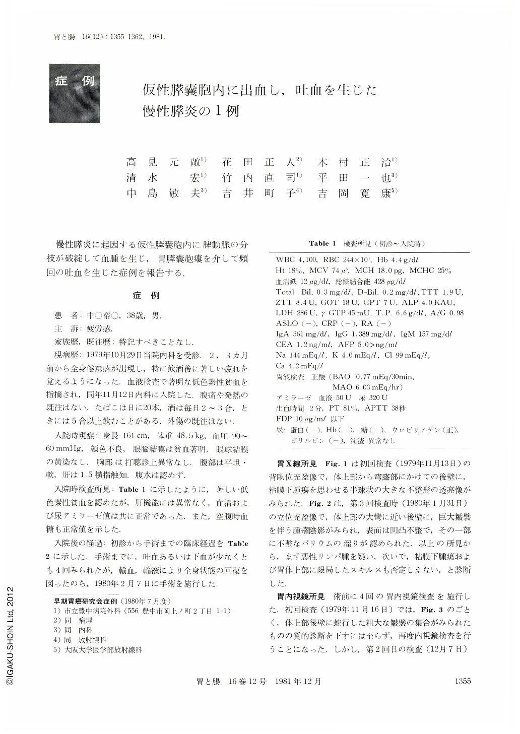
慢性膵炎に起因する仮性膵嚢胞内に脾動脈の分枝が破綻して血腫を生じ,胃膵囊胞瘻を介して頻回の吐血を生じた症例を報告する..
A case of pancreatic pseudocyst causing massive gastric bleeding is reported, and the diagnostic importance of celiac angiography for this condition is emphasized.
The patient, a 38-year-old man, was admitted to the hospital because of general fatigue of several months' duration. A heavy alcoholic history was obtained, but there were no previous episodes suggestive of pancreatitis such as upper abdominal pain. He had severe hypochromic anemia, and his serum amylase was normal. Routine upper G-I series showed abnormal folds over the posterior wall of the upper fundic region accompanied by signs of extrinsic compression. During the course of endoscopic examination, he developed suddenly massive gastric bleeding of obscure origin. To determine the source of hemorrhage, celiac angiography was subsequently performed. This showed a 3.7 by 2.8 cm, rounded, homogenous opacification in the tail of the pancreas, which was most intensely demonstrated at the venous phase. Computed abdominal scan showed enlargement of the tail of the pancreas suggestive of a pancreatic tumor. Because of recurring gastric bleeding, laparotomy was done, and en bloc resection of the involed organs including the tail of the pancreas, stomach, and spleen was performed. Pathologic examination showed histological evidence of chronic pancreatitis and a blood-filled pseudocyst in the tail of the pancreas, which created a gastrocystic fistula. In addition, branches of the splenic artery as well as a large pancreatic duct were found to communicate with the pseudocyst.

Copyright © 1981, Igaku-Shoin Ltd. All rights reserved.


