Japanese
English
- 有料閲覧
- Abstract 文献概要
- 1ページ目 Look Inside
- サイト内被引用 Cited by
虫垂に発生したCrohn病は,欧米では相次いで報告されている1~11)が,わが国では,このような限局性の病変をCrohn病と診断することを疑問視する向きもあり,虫垂Crohn病の報告はほとんどみられない.
われわれは最近,虫垂ならびに盲腸に限局したCrohn病と考えられる症例を経験したので,その詳細を報告し,併せて文献的考察を加えた.
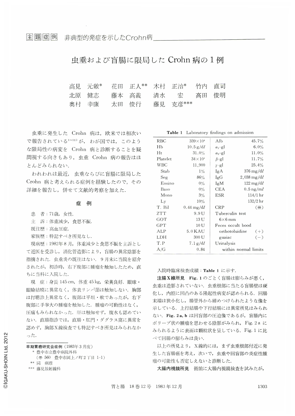
A case of Crohn's disease confined to the appendix and adjacent cecum is presented. The Patient, a 70-year-old woman, was admitted to the hospital with complaints of anorexia and loss of body weight of two months' duration. Physical examination showed a tender mass in the right lower quadrant of the abdomen. Mantoux test was questionably positive with a 6 mm area of redness. Other laburitory data, including chest x-rays, were all within normal limits. Barium enema study revealed a polypoid lesion in the cecum and possibly extrinsic narrowing of the terminal ileum. An attempt at colonofiberscopy was unsuccessful. A neoplasm of the cecum could not be ruled out so that an exploratory lapurotumy was done. At operation, the ileocecal region was markedly thickened and adherent by inflammation. Intraoperative colonoscopy revealed a cobblestone-like mucosal lesion in the cecum without ulceration of the mucosa. A right hemicolectomy was performed with an end-to-end anastomosis. The gross resected specimen, measuring 30 cm in length, showed a thickened cecal wall with marked pericolic fibrosis. The appendix was embedded within the cecal inflammatory mass. The mucosal surface of the cecum had a conglomerated, polypoid appearance limited near to the orifice of the appendix, but no mucosal lesions were found in the terminal ileum. Histological examination showed transmural inflammation with fibrosis of the cecal wall and finger-like inflammatory polyps near the orifice of the appendix. Multiple, noncaseating, epithelioid granulomas, some of which containing Langhans' type giant cells, were seen mainly in the lamina propria and submucosa of the appendix and the cecal polypoid regions. There were also superficial and deep fissuring ulcers and intramural abscesses. The terminal ileum and the rest of colon were free of granulomatous involveInent.
Since the first report by Meyerding in 1953, approximately 60 cases of primary appendiceal Crohn's disease have been reported in the English-language literature. These cases include those of Crohn's disease that were limited to the appendix, or with continuity to the adjacent cecum. Our search of the literature in Japan disclosed no similar reports. However, there are three well-documented cases of typhlitis exhibiting histologically features of Crohn's had a previous history of appendectomy one to three years before, but the histology of these resected appendices was not described in any detail. Nevertheless, we were impressed by striking radiographic, endoscopic, as well as pathologic similarities between these cases and ours, as far as the cecal lesion is concerned.
Copyright © 1983, Igaku-Shoin Ltd. All rights reserved.