Japanese
English
- 有料閲覧
- Abstract 文献概要
- 1ページ目 Look Inside
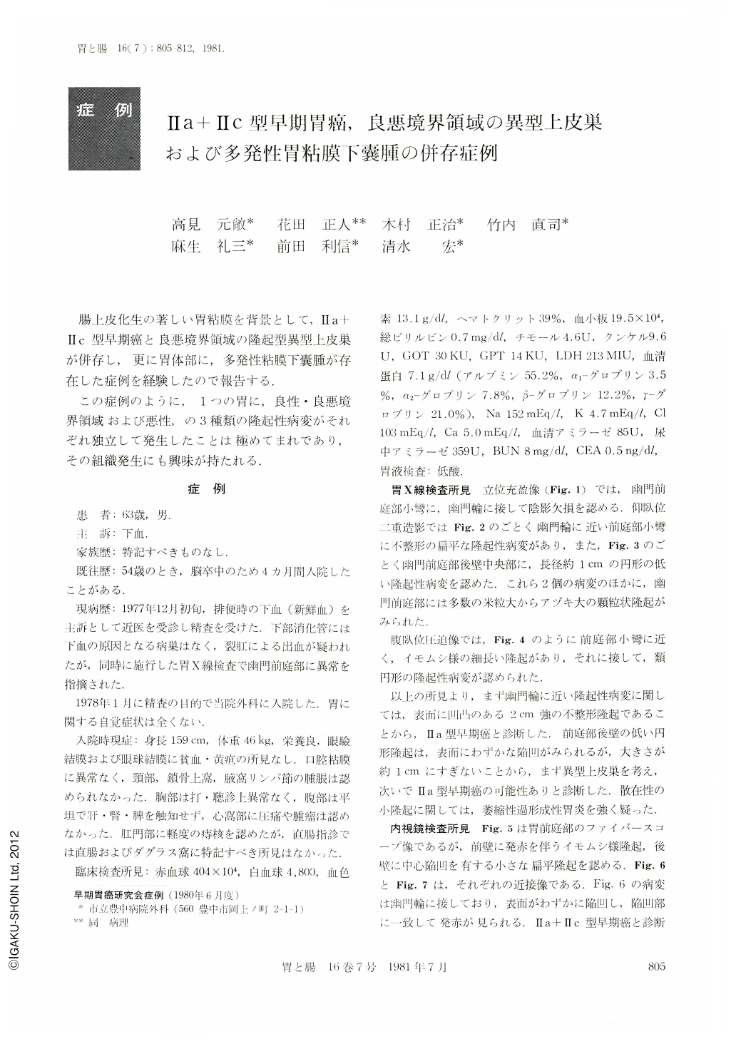
腸上皮化生の著しい胃粘膜を背景として,Ⅱa+Ⅱc型早期癌と良悪境界領域の隆起型異型上皮巣が併存し,更に胃体部に,多発性粘膜下嚢腫が存在した症例を経験したので報告する.
この症例のように,1つの胃に,良性・良悪境界領域および悪性,の3種類の隆起性病変がそれぞれ独立して発生したことは極めてまれであり,その組織発生にも興味が持たれる.
A 63-year-old man, who had had no related gastric symptom, was found to have several isolated elevated lesions in the stomach by roentgenographic and endoscopic examinations: these included two lesions in the antrum resembling early gastric carcinoma of type Ⅱa+Ⅱc and a few submucosal cystic lesions in the corpus. Besides these, the antral mucosa was coarsely granular suggestive of atrophic hyperplastic gastritis. Biopsies taken from the former two lesions were read as Group Ⅴ (cancer) and Group Ⅲ (border line lesion), respectively. Pathologic examination of the resected specimen confirmed an early adenocarcinoma limited to the mucosa (2.3×1.9cm in size) and an atypical epithelium (1.4×1.0cm in size), which were present separately. The submucosal lesions found in the corpus showed multiple submucosal cysts composed of hyperplastic foveolar and pyloric-gland-type of epithelium containing some fundic cells. Although there was marked diffuse intestinal metaplasia of both pyloric and fundic mucosae, no such changes were observed in the submucosal glands. In one of these lesions a central pit connecting the foveolar epithelium of the overlying mucosa and that of submucosal cysts was demonstrated. The coexistence of marked diffuse intestinal metaplasia, submucosal cysts, atypical epithelium, and flank adenocarcinoma in our case appears to go well with the view that these lesions probably have a common etiopathogenetic background in their development, although cases with such complex gastric lesions are extremely rare.

Copyright © 1981, Igaku-Shoin Ltd. All rights reserved.


