Japanese
English
- 有料閲覧
- Abstract 文献概要
- 1ページ目 Look Inside
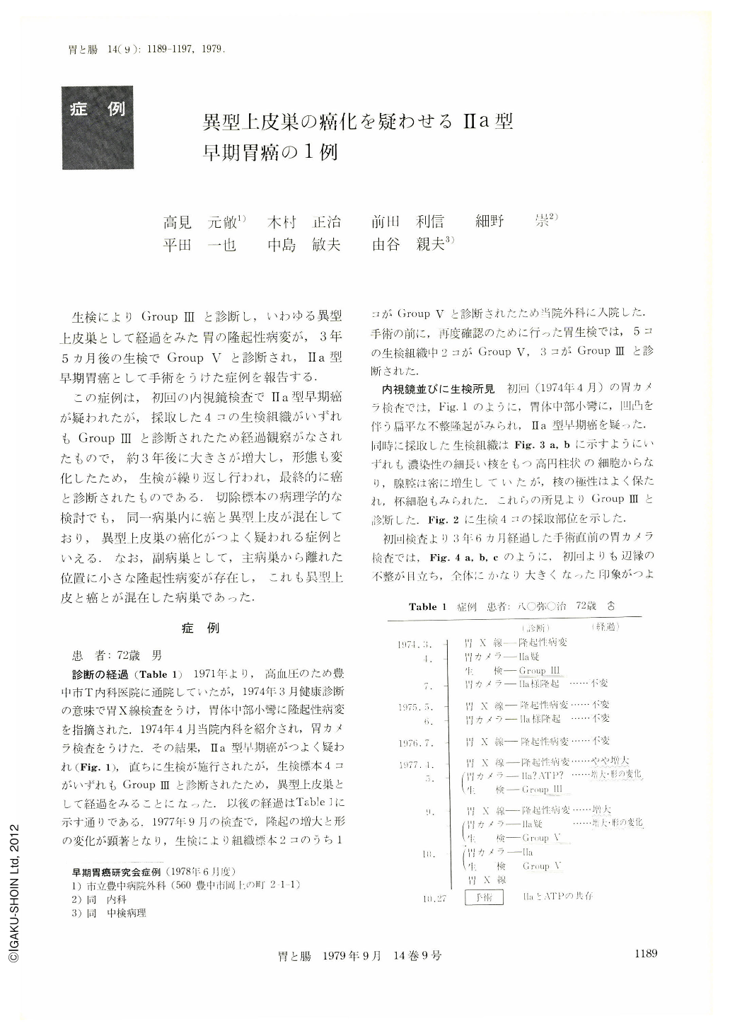
生検によりGroupⅢと診断し,いわゆる異型上皮巣として経過をみた胃の隆起性病変が,3年5カ月後の生検でGroupⅤと診断され,Ⅱa型早期胃癌として手術をうけた症例を報告する.
この症例は,初回の内視鏡検査でⅡa型早期癌が疑われたが,採取した4コの生検組織がいずれもGroupⅢと診断されたため経過観察がなされたもので,約3年後に大きさが増大し,形態も変化したため,生検が繰り返し行われ,最終的に癌と診断されたものである.切除標本の病理学的な検討でも,同一病巣内に癌と異型上皮が混在しており,異型上皮巣の癌化がつよく疑われる症例といえる.なお,副病巣として,主病巣から離れた位置に小さな隆起性病変が存在し,これも異型上皮と癌とが混在した病巣であった.
A man 72 years of age, free from any symptoms related to the digestive tract, had undergone examination of the stomach in April 1974, when a shadow defect had been noticed in the midbody. Subsequent gastroscopy revealed a flat elevated lesion in the midbody. Gastric biopsy led us to a diagnosis of Group Ⅲ, namely a border line lesion, so we kept on following him up.
About three years after the first examination, X-ray and endoscopic examination showed that the elevated lesion slightly enlarged and changed in shape. Gastric biopsy in Sept. 1977 revealed it to be malignant (Group Ⅴ), and the patient was operated on for early gastric cancer.
Resected stomach showed a sessile polypoid lesion in the body, 2.8×2.2 cm in size. Histopathologically it showed tubular adenocarcinoma accompanied with atypical epithelium, which was confined to the mucosa.
In addition there was a small elevated lesion in the antrum, which showed an atypical epithelium partly coexisted with cancerous area. This small lesion had been overlooked before operation.
We concluded that this case was a very rare case which veritably showed malignant transformation of an atypical epithelium.

Copyright © 1979, Igaku-Shoin Ltd. All rights reserved.


