Japanese
English
- 有料閲覧
- Abstract 文献概要
- 1ページ目 Look Inside
“早期”胃悪性リンパ腫の報告例は相当数にのぼり,その臨床的ならびに形態学的特徴も明らかとなってきた.われわれも病変発見時より胃悪性リンパ腫を疑い,8カ月の経過観察で術前に確診しえた症例を経験したので報告する.
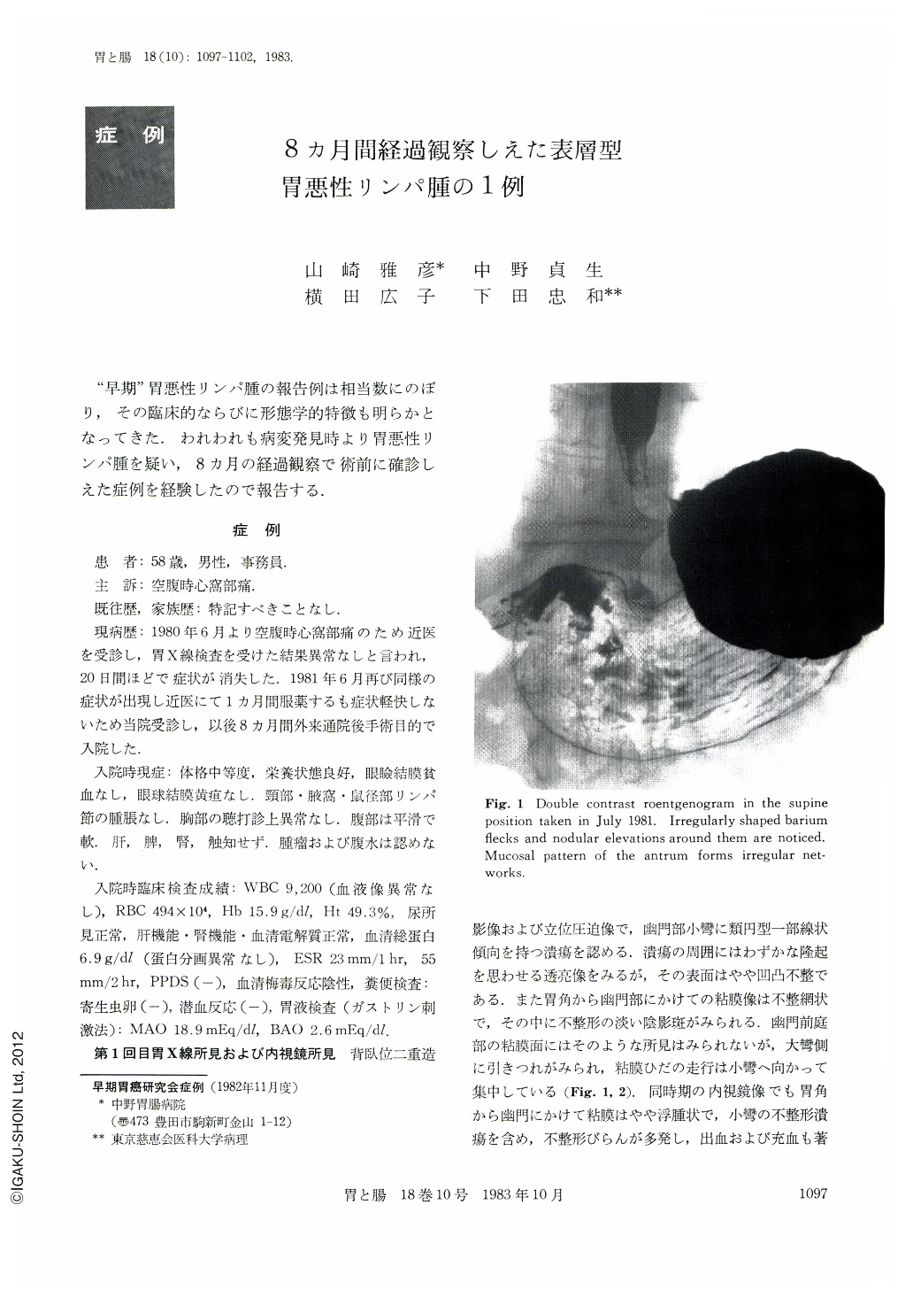
The patient, a 58 year-old man, visited our hospital on July 1, 1981, with a chief complaint of hunger epigastralgia that continued for a month. Physical examination and study of the urine, feces and blood were normal. On the other hand, x-ray examination of the stomach showed an ovoid ulcer and multiple erosions from the angle down to the antrum. The mucosal surface around them was irregularly granular. The areas between each granular showed relatively distinct grooves. The border between them and normal mucosa was not clear. Endoscopy also revealed multiple ulcers, nodular elevations around them and multiple bleeding spots. The lesion as a whole was edematous. These findings led us to suspect malignant lymphoma or reactive lymphoreticular hyperplasia. During the eight months' follow-up ulcers healed and appeared repeatedly, but nodular elevations around them became remarkable. The fourth biopsy of the lesion revealed histologic findings of malignant lymphoma, so that subtotal gastrectomy was performed. The postoperative course was uneventful. Resected specimen of the stomach showed macroscopically finely or coarsely granular mucosal surface with converging folds and an active ulcer on the antrum. The border between the lesion and the normal mucosa was again not clear. These findings obviously differed from those of Ⅱc type early gastric cancer. Histologically the lesion was malignant lymphoma of diffusely mixed cellular type. Tumor cells invaded only the mucosa.

Copyright © 1983, Igaku-Shoin Ltd. All rights reserved.


