Japanese
English
- 有料閲覧
- Abstract 文献概要
- 1ページ目 Look Inside
胃悪性リンパ腫の全身性に転移したと考えられる症例を経験したので,X線,内視鏡所見および病理学的所見につき供覧する.
症 例
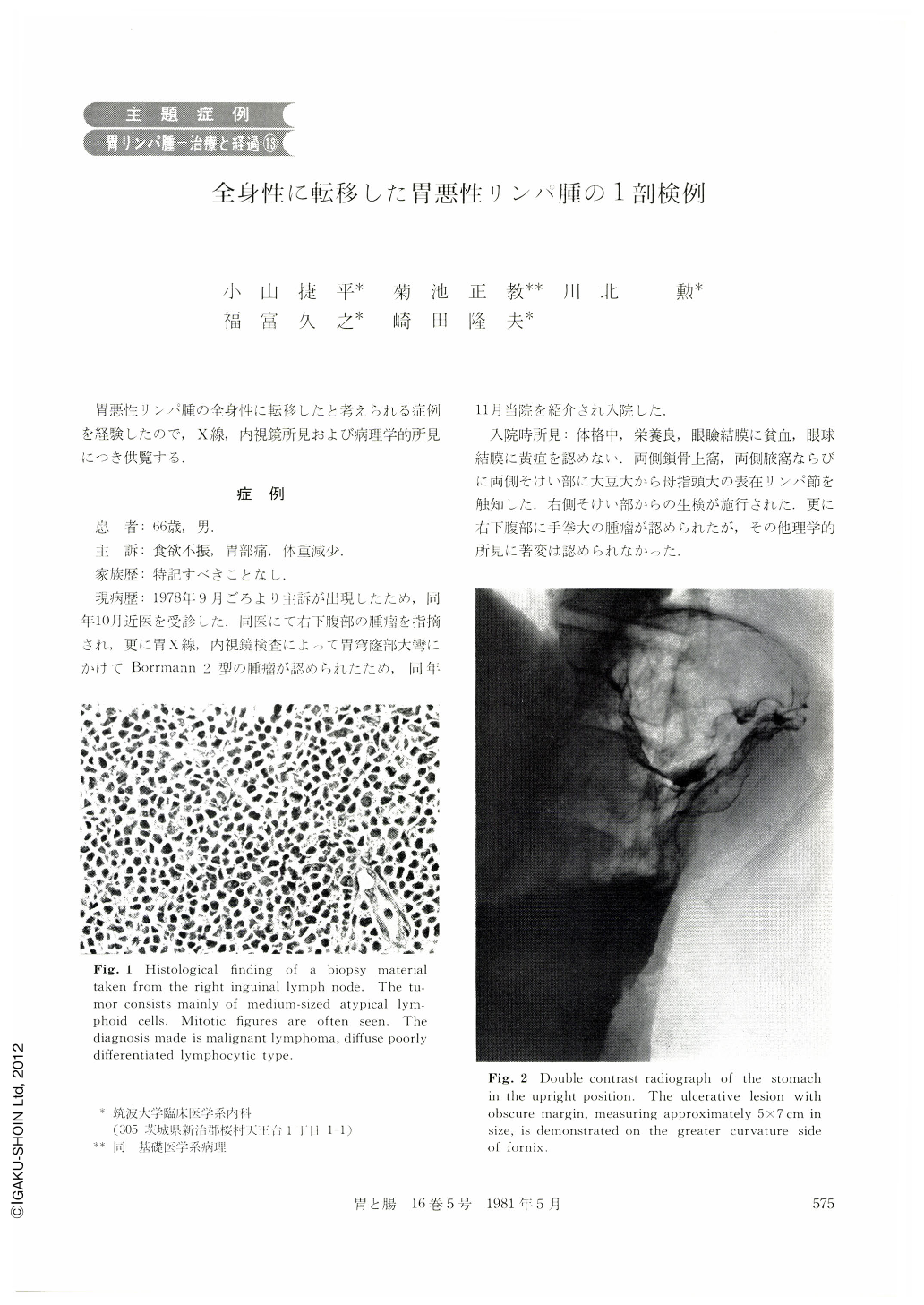
患 者:66歳,男.
主 訴:食欲不振,胃部痛,体重減少.
家族歴:特記すべきことなし.
現病歴:1978年9月ごろより主訴が出現したため,同年10月近医を受診した.同医にて右下腹部の腫瘤を指摘され,更に胃X線,内視鏡検査によりて胃穹窿大彎にかけてBorrmann 2型の腫瘤が認められたため,同年11月当院を紹介され入院した.
A 66-year-old man was admitted to our hospital complaining of appetite loss, epigastric pain and weight loss which had appeared since two months before.
The radiological examination of the upper gastrointestinal tract revealed an ulcerative lesion with obscure margin, measuring approximately 5×7 cm, in the fornix of the greater curvature side. Endoscopic examination disclosed an ulcerative lesion with white coat and some bleeding spots. The surface and outer limit of the raised margin of this lesion were too smooth and seemed to be covered with normal epithelium. Furthermore, no worm-eating appearance or fusion at the tips of the covering mucosal folds were discovered. The lesion had some findings of gastric mucosal tumor. Biopsy materials taken from the stomach and inguinal lymph node were diagnosed histologically as malignant lymphoma (diffuse, poorly differentiated lymphocytic type, classified by Rappaport).
Since wide-spread metastasis of malignant lymphoma originated from the stomach was ascertained, chemotherapy was (Vincristine, Cyclophosphamide, 6-Mercaptopurine, Prednisolone) started on the 14th day. After that, general condition of the patient had improved and man's fist sized tumor of the lower ahdomen became smaller. Unfortunately, pulling out tooth because of toothache on the 37th day caused a sepsis. The patient died on the 56th day.
Necropsy was performed. Macroscopic examination of the necropsied stomach revealed two malignant lymphomas. A 5×3 cm ulcer with smoothly raised margin was observed as Borrmann 2 type lesion and located in the posterior wall of the fornix. Histologically, the tumor cells of the fornix infiltrated diffusely in all layers of the gastric wall. The size of malignant lymphoma infiltration was 7×5 cm. A small elevated lesion, measuring 1.5 cm in diameters with tiny erosion was located at the lower portion of the corpus. The tumor cells were limited in the lamina propria mucosae and submucosa. Hematogeneous infiltration of malignant lymphoma was observed in the liver, lungs, spleen, kidney, heart, tongue, esophagas, ileum, peritoneum, epididymis, prostate and bone-marrow. Lymphogenous infiltration was seen in abdominal para-aortal, mesenterial, retroperitoneal, liver hilar, lung hilar, carinal, para-tracheal and inguinal nodes.
Finally, the case had systemic lymphosarcomatous state and the immediate cause of death was pulmonary bleeding by tumor cells infiltration.
Copyright © 1981, Igaku-Shoin Ltd. All rights reserved.