Japanese
English
- 有料閲覧
- Abstract 文献概要
- 1ページ目 Look Inside
本症例は,術前胃体中部後壁にBorrmann2型進行癌と左鎖骨上窩リンパ節転移が証明され,開腹所見で原発腫瘍の膵尾部への浸潤とN1,2(+)と判定されて単開腹にとどめ,術後少量のFAMT療法を行ったのみで退院したが,退院後急速に全身状態の改善をみるとともに,原発巣ならびにリンパ節転移も縮小,消失して,9年半を経過し,今日健在している.
A 62 year-old man with epigastric dull pain and sense of fullness was diagnosed as having gastric carcinoma by mass survey.
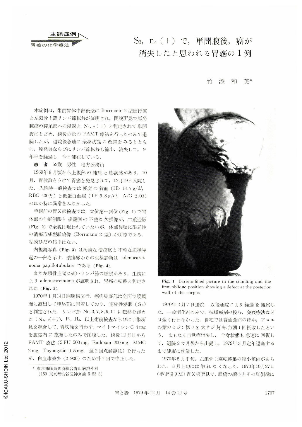
X-ray examination on admission revealed a Borrmann type 2 carcinoma on the posterior wall of the upper corpus (Figs. 1, 2). Endoscopic study showed a dirty irregular ulceration surrounded by clearly outlined, high marginal wall (Fig. 3). Adenocarcinoma papillotubulare was proved by endoscopic biopsy (Fig. 4).
One of the swollen lymphnodes in the left supraclavicular fossa was also biopsied proving adenocarcinoma and it was interpreted as a metastasis from the gastric cancer.
A laparotomy was done in January, 1970. The resection was not performed since the gastric tumor of the posterior wall of the middle corpus directly invaded into the tail of the pancreas and lymphnode metastasis in the paraaortic area was apparent.
Anti-cancer chemotherapy was started on the 12th postoperative day with a combination of 5-FU 500mg, Endoxan 200mg, Mitomycin C 2 mg, and Toyomycin O.5mg by intravenous drip infusion twice a week, but it was discontinued after seven courses because of decrease of leucocyte.
The patient was followed in the outpatient clinic without any other chemotherapy or immunotherapy.
Seven months postoperatively, swelling of the lymphnodes of the left supraclavicular fossa disappeared. Nine month after the operation a marked decrease in the size of the gastric tumor was noted by x-ray. One year and 7 months postoperatively, an X-ray study showed complete disappearance of the Borrmann type 2 carcinoma leaving only a finding simulating a scar of peptic ulcer (Figs. 6, 7). The X-ray finding has been similar thereafter by examinations done once every year. An endoscopic examination, 6 years and 5 months postoperatively, showed a slightly discolored area surrounded by puckering folds and some tips of the converging folds appeared to Show small nodulation (Fig. 8). Endoscopic biopsy specimens taken from the area were all regenerating mucosa with no evidence of carcinoma (Fig. 10). An X-ray study, 8 years and 11 months postoperatively, showing only converging folds similar to the previous ones, is demonstrated in Fig 9.
The patient is alive and well at present, 9 years and 9 months after the laparotomy.
The main factor for the rather quick and continued disapperance of advanced carcinoma in this patient is unknown, Although the dosage of the anti-cancer chemotherapy was quite small, it might have activated or promoted an anti-tumor ability of the host.

Copyright © 1979, Igaku-Shoin Ltd. All rights reserved.


