Japanese
English
- 有料閲覧
- Abstract 文献概要
- 1ページ目 Look Inside
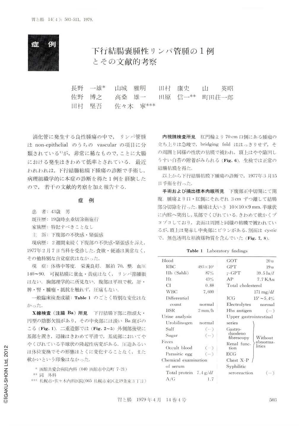
消化管に発生する良性腫瘍の中で,リンパ管腫はnon-epithelialのうちのvascularの項目に分類されている1)が,非常に稀なもので,ことに大腸における発生はきわめて低率とされている.最近われわれは,下行結腸粘膜下腫瘍の診断で手術し,病理組織学的に本症の診断を得た1例を経験したので,若干の文献的考察を加え報告する.
Out of the benign tumors that develop in the digestive tract, lymphangioma is classified as the “vascular type” in the category of non-epithelial tumors. However, it is a very rare lesion, and especially its occurrence in the large bowel is accepted as very low. We have recently encountered a case which was operated on under the diagnosis of submucosal tumor of the descending colon and which was histopathologically diagnosed as lymphangioma; and the findings in this case are presented hereunder together with a discussion with reference to literature data.
The patient, a 43-year-old man, with no other past history than appendectomy at the age of 19, came to the author's hospital on February 7, 1977 for complaints of dysphoria and tense feeling in the lower abdominal region persisting for two weeks. The findings by physical examinations were normal, and laboratory findings were in no way abnormal. Barium enema study revealed that there was a round filling defect of a finger-tip size in the distal portion of the descending colon with a thin barium spot remaining in its center. Double contrast roentgenography disclosed a semispheric, elevated lesion, which was based on the latero-posterior wall of the distal portion of the descending colon, with a very smooth margin and with a slight constriction in its base. Neither compression nor change of body position caused the lesion to change its shape, and it gave no impression of a soft lesion. Colonofiberscupy showed that the tumor located 70 cm oral to the anus uprose sharply; the bridging folds were not clear; the lesion was coated with the mucous membrane similar in nature to that in the adjacent area; its tip was slightly depressed with a white coat attached there. Biopsy showed the normal colonic mucoua.
From these findings, the patient was operated on March 15, 1977 under the diagnosis of submucosal tumor of the descending colon. Partial colectomy was performed between the parts 3 cm oral to and 3 cm anal to the tumor. The tumor, measuring 10×10×9 mm, protruded into the lumen of the colon in a semispheric form, with a constriction at its base. It was very soft, and spongy; its surface was covered with a mucous membrane similar to that in the adjacent area, but its tip was red with an erosion in the center. The cross section was cystica nd contained a colorless, clear, mucous-like substance.
Histologically, there were many cystoma-like structures varying in size chiefly in the submucosa, some of them involving the lamina propria mucosae, and they were filled with a secretion which stained lightly with eosin. The wall of the cystoma-like structures consisted of monolayer, flat endothelial cells. These findings led to the diagnosis of “Lymphangioma cystoides”.

Copyright © 1979, Igaku-Shoin Ltd. All rights reserved.


