Japanese
English
- 有料閲覧
- Abstract 文献概要
- 1ページ目 Look Inside
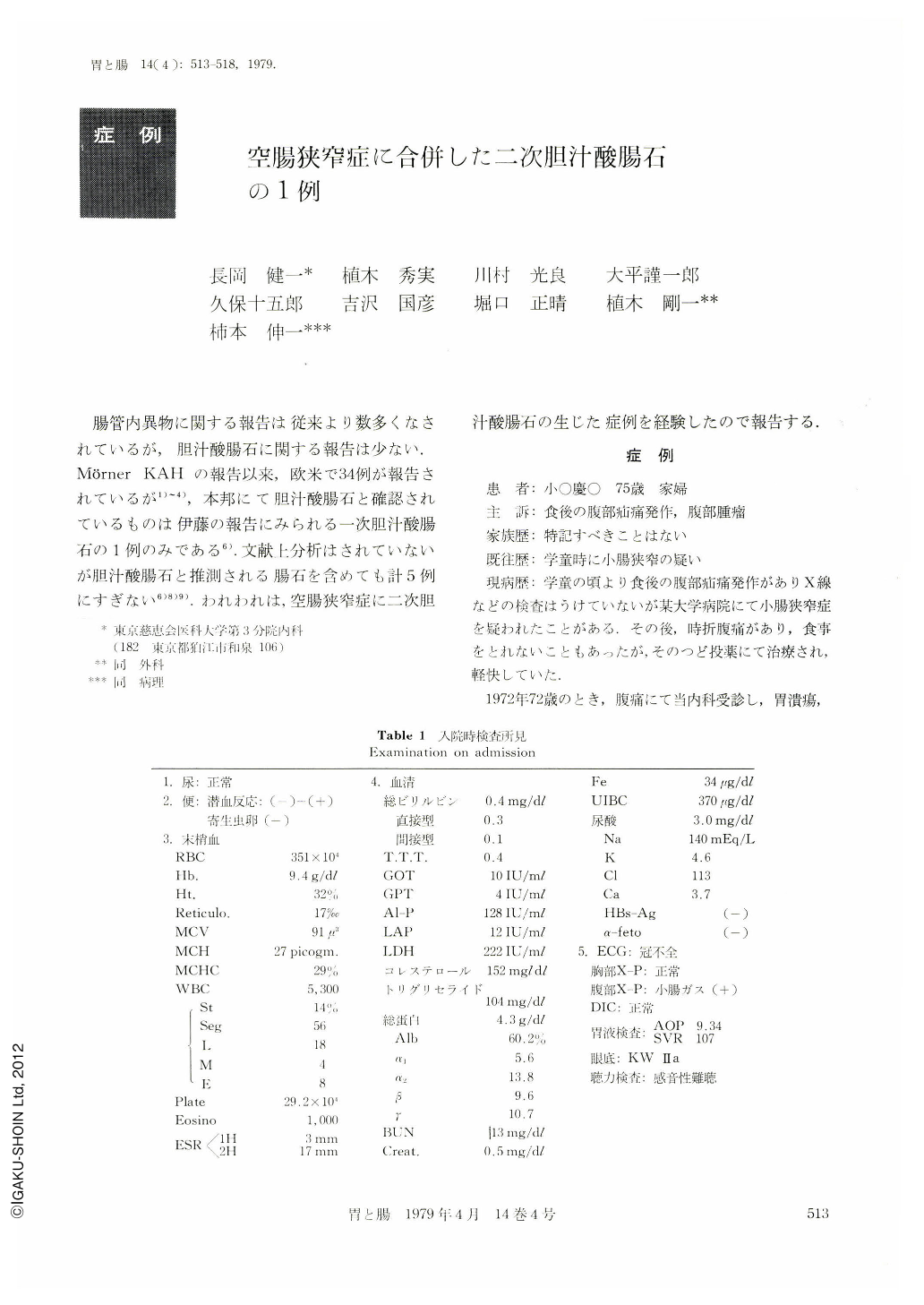
腸管内異物に関する報告は従来より数多くなされているが,胆汁酸腸石に関する報告は少ない.Mörner KAHの報告以来,欧米で34例が報告されているが1)~4),本邦にて胆汁酸腸石と確認されているものは伊藤の報告にみられる一次胆汁酸腸石の1例のみである6).文献上分析はされていないが胆汁酸腸石と推測される腸石を含めても計5例にすぎない6)8)9).われわれは,空腸狭窄症に二次胆汁酸腸石の生じた症例を経験したので報告する.
Seventy-five years old Japanese woman complained of colicky abdominal pain after meals. Physical examination demonstrated 6.5 cm hard large tumor in the right upper quadrant. During her childhood, she had experienced episodes of intermittent abdominal pain.
Fluoroscopy of the digestive tract after admission showed persistent filling defect suggestive of a stone possibly in the jejunal loop.
At laparoscopy, we found 6.5×6.5 cm yellow enterolith located 200 cm proximal to the ileocecal valve. Just anal site of enterolith showed marked narrowing, allowing only a finger tip to pass through. Histological examination of narrowing part demonstrated intact mucous layer, nerve and blood vessel, suggestive of congenital stenosis. UV spectrum analysis showed that the enterolith consisted of mainly deoxycholic acid and a small amount of bilirubin. The deoxycholic acid was of free type.
Bile acid enterolith is rare, and only 35 cases have been reported in the world literature. In Japan, this is the second case of bile acid enterolith and the first case of secondary bile acid enterolith.

Copyright © 1979, Igaku-Shoin Ltd. All rights reserved.


