Japanese
English
- 有料閲覧
- Abstract 文献概要
- 1ページ目 Look Inside
- サイト内被引用 Cited by
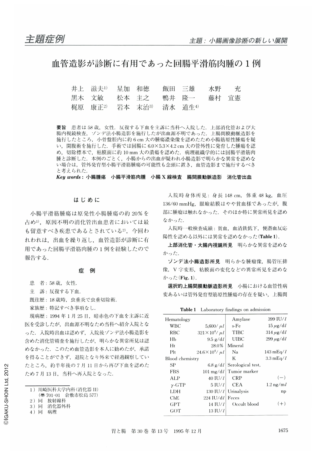
要旨 患者は58歳,女性.反復する下血を主訴に当科へ入院した.上部消化管および大腸内視鏡検査,ゾンデ法小腸造影を施行したが出血源不明であった.上腸間膜動脈造影を施行したところ,小骨盤腔内に約6cm大の腫瘍濃染像を認めたため小腸筋原性腫瘍を疑い,開腹術を施行した.手術では回腸に6.0×5.3×4.2cm大の管外性に発育した腫瘍を認め,切除標本で,粘膜面に約10mm大の潰瘍を認めた.病理組織学的には回腸平滑筋肉腫と診断した.本例のごとく,小腸からの出血が疑われ小腸造影で明らかな異常を認めない場合は,管外発育型小腸平滑筋腫瘍の可能性も念頭に置き,血管造影まで施行するべきと考えられた.
A 58-year-old woman was admitted to our division due to repeated hematochizia. Although upper gastrointestinal endoscopic, colonoscopic and double-contrast small intestinal radiologic examinations failed to identify any lesions that could be a source of hematochizia, superior mesenteric arteriography revealed an ovalshaped hypervascular mass in the pelvic cavity. The tumor was also identifed as a large, homogenous mass by CT and MRI scans. In addition, it was demonstrated as a hypoechoic mass by the endoscopic ultrasonographic examination scanned through the rectal wall. The patient was treated surgically, and the histologic examination of the resected specimen showed leiomyosarcoma developed from the ileal wall. Retrospective review of the x-ray films of the small intestine showed a minimal eccentric deformity which seemed to be adjacent to the tumor in the ileum. Barium examination of the small intestine seemed to be insufficient to diagnose leiomyosarcoma of the small intestine, because it grew extraluminally with minimal intraluminal growth. Our case suggests that angiography may be useful for the diagnosis of small intestinal tumors.

Copyright © 1995, Igaku-Shoin Ltd. All rights reserved.


