Japanese
English
- 有料閲覧
- Abstract 文献概要
- 1ページ目 Look Inside
近年膵疾患の診断には,十二指腸ファイバースコープによる逆行性膵管造影検査および直接膵液採取による細胞診が行なわれるようになり,その診断率は向上してきた.しかし,膵管造影所見のみでは慢性膵炎と膵癌とは鑑別困難なことも少なくない.また,膵液細胞診は膵液中に常に一定の数の新鮮な癌細胞の遊出を条件とするため,膵体尾部癌の診断には限界がある.
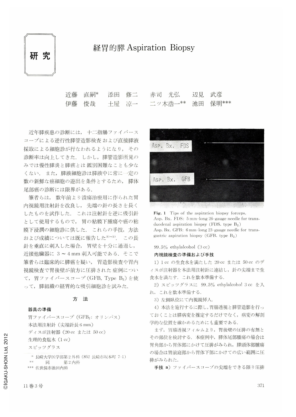
筆者らは,数年前より潰瘍治療用に作られた胃内視鏡用注射針を改良し,先端の針の長さを長くしたものを試作した.これは注射針を逆に吸引針として使用するもので,胃の粘膜下腫瘍や癌の粘膜下浸潤の細胞診に供した.これらの手技,方法および成績については既に報告した3)~5).この長針を輩直に刺入した場合,胃壁を十分に通過し,近接他臓器に3~4mm刺入可能である.そこで筆者らは臨床的に膵癌を疑い,胃造影検査や胃内視鏡検査で胃後壁が前方に圧排された症例について,胃ファイバースコープ(GFB,Type B2)を使って,膵組織の経胃的な吸引細胞診を試みた.
The most accurate method to establish definite diagnosis of pancreatic cancer may be incisional biopsy and aspiration biopsy under direct vision of the pancreas upon laparotomy. However, t is not reasonable to perform exploratory laparotomy for all patients of suspected pancreatic cancer. Hence, various non-surgical methods for its diagnosis have been devised. These methods consist mostly in cytological examination of, for example, bile sampled at the time of percutaneous transhepatic cholangiography, duodenal juice obtained at the time of pancreozyminsecretin test, and pancreatic juice sampled after endoscopic pancreatography using duodenal fiberscope, or transduodenal pancreatic aspiration biopsy6) which we devised.
These methods have the advantage of being available at the time of other tests and have their own significance. However, all these methods are suitable for the diagnosis of cancer of the head of the pancreas but not of the body and tail of the pancreas. We devised an easier and yet a most convenient method for the diagnosis of lesion in the body and tail of the pancreas that had been the most difficult region for diagnosis.
In endoscopic exmination by means of gastrofiberscope, the body and tail of the pancreas can be recognized as an elevation on the lesser curvature of the body of the stomach. This elevation increases its size according to the size of the tumor of the body and tail of the pancreas and it usually appeare as an irregularly uneven protrusion having a bridging fold on the greater curvature. If such a protrusion is observed at endoscopic examination, pancreatic tissue is collected by several aspiration biopsies using an aspiration fine needle which we have devised. The specimen is divided into the smears and the cellblock for cytological examination.
Thus, we checked the accuracy of this new method in the cases wherein the posterior wall of the body stomach was seemingly oppressed by the pancreatic lesion. As a result, it was found that the malignant cells had been collected 100 per cent in malignant tumors of the pancreas that opperessed the posterior wall of the stomach. Moreover, this method was confirmed to be safe without any complications such as hemorrhage.
Copyright © 1976, Igaku-Shoin Ltd. All rights reserved.