Japanese
English
- 有料閲覧
- Abstract 文献概要
- 1ページ目 Look Inside
胃内視鏡検査は,X線検査と表裏一体をなすものとして,胃疾患診断上不可欠のものとなって来た.しかし胃粘膜下の腫瘍の質的診断は,直視下胃生検の結果を持ってしてもなお困難を要する所である.
筆者らは最近,胃幽門部前庭に臍状陥凹を有する典型的と思われる胃壁内迷入膵の症例を経験したので報告したい.
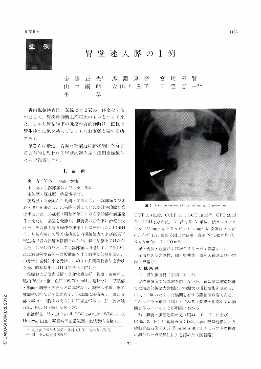
The patient, a woman 39 years of age, was admitted here on account of epigastric and right hypochondrial pain. No pathological finding was noticed in the general examination. Double contrast radiograph of the stomach revealed a circular shadow the size of a forefinger-tip on the greater curvature near the posterior wall of the antrum. Frontal compression of this region also disclosed a filling defect with central depression. Concomitantly, many stones were seen on the cholecysto-cholangiograph. Endoscopically, the above lesion was observed as a small, nipple-like structure projecting from the surrounding mucosa. The mucosal surface was free from discoloration, bleeding or edema, nor was there any bridging fold to be seen. Biopsy under direct vision afforded no diagnostic information for this lesion. The operation was done under the combined diagnosis of a submucosal tumor, cholelithiasis, choledocholithiasis and chronic cholecysto-cholangitis. In the resected stomach, a nipple-like tumor, measuring 11.7×11.3×7.4mm, was found in the pyloric antrum 1.5cm oral from the pyloric ring. Under this papilla, a thumb-tip-sized firm mass could be felt within the wall of the stomach. Histopathologically, a part of the tunica propria in the tip of the lesion had been concaved and depressed into the submucosa. The exocrine portion of the heterotopic pancreatic tissue without ducts was also revealed in the tunica propria together with the Brunner's glands. The exocrine part without Langerhans' islet was also seen in the submucosa. Beneath the nipple-like tumor was recognized “adenomyosis” of the muscularis. The orifice of the ducts in the center of the papilla could not be identified, however.

Copyright © 1971, Igaku-Shoin Ltd. All rights reserved.


