Japanese
English
- 有料閲覧
- Abstract 文献概要
- 1ページ目 Look Inside
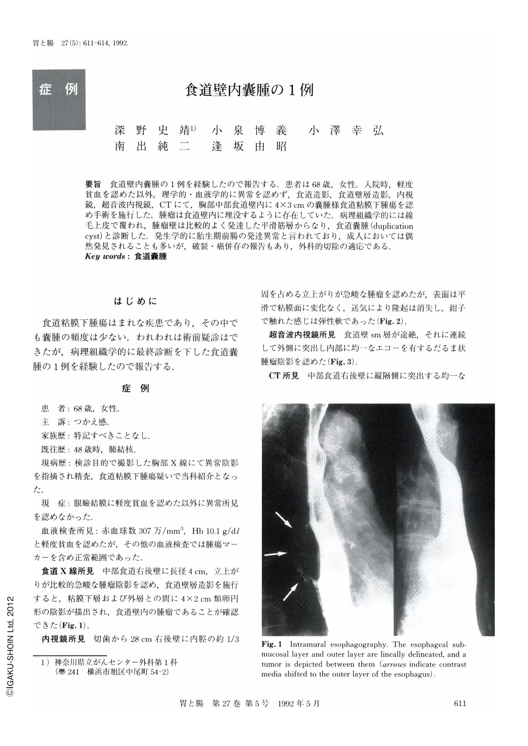
要旨 食道壁内囊腫の1例を経験したので報告する.患者は68歳,女性.入院時,軽度貧血を認めた以外,理学的・血液学的に異常を認めず,食道造影,食道壁層造影,内視鏡,超音波内視鏡,CTにて,胸部中部食道壁内に4×3cmの囊腫様食道粘膜下腫瘍を認め手術を施行した.腫瘤は食道壁内に埋没するように存在していた.病理組織学的には線毛上皮で覆われ,腫瘤壁は比較的よく発達した平滑筋層からなり,食道囊腫(duphcationcyst)と診断した.発生学的に胎生期前腸の発達異常と言われており,成人においては偶然発見されることも多いが,破裂・癌併存の報告もあり,外科的切除の適応である.
A case of intramural esphageal cyst was encountered and is reported here. The patient was a 68-year-old female. Physical examination and hematology on admission revealed no abnormalities except for slight anemia. A cystic submucosal tumor 4×3 cm in size was observed intramurally in the middle thoracic esophagus by esophagography, intramural esophagography, endoscopy, endoscopic ultrasonography and CT, and was surgically removed. The tumor appeared as if it were buried in the esophageal wall. Histopathologically, it was covered with ciliated epithelia, and the tumor wall consisted of relatively well developed smooth muscle layers. It was diagnosed as a duplication cyst. Embryologically, it is said to be a developmental anomaly of the foregut. In adults, it is discovereda, unexpectedly in most cases, but there are reports of rupture or complications with cancer, so surgical resection is indicated.

Copyright © 1992, Igaku-Shoin Ltd. All rights reserved.


