Japanese
English
- 有料閲覧
- Abstract 文献概要
- 1ページ目 Look Inside
- サイト内被引用 Cited by
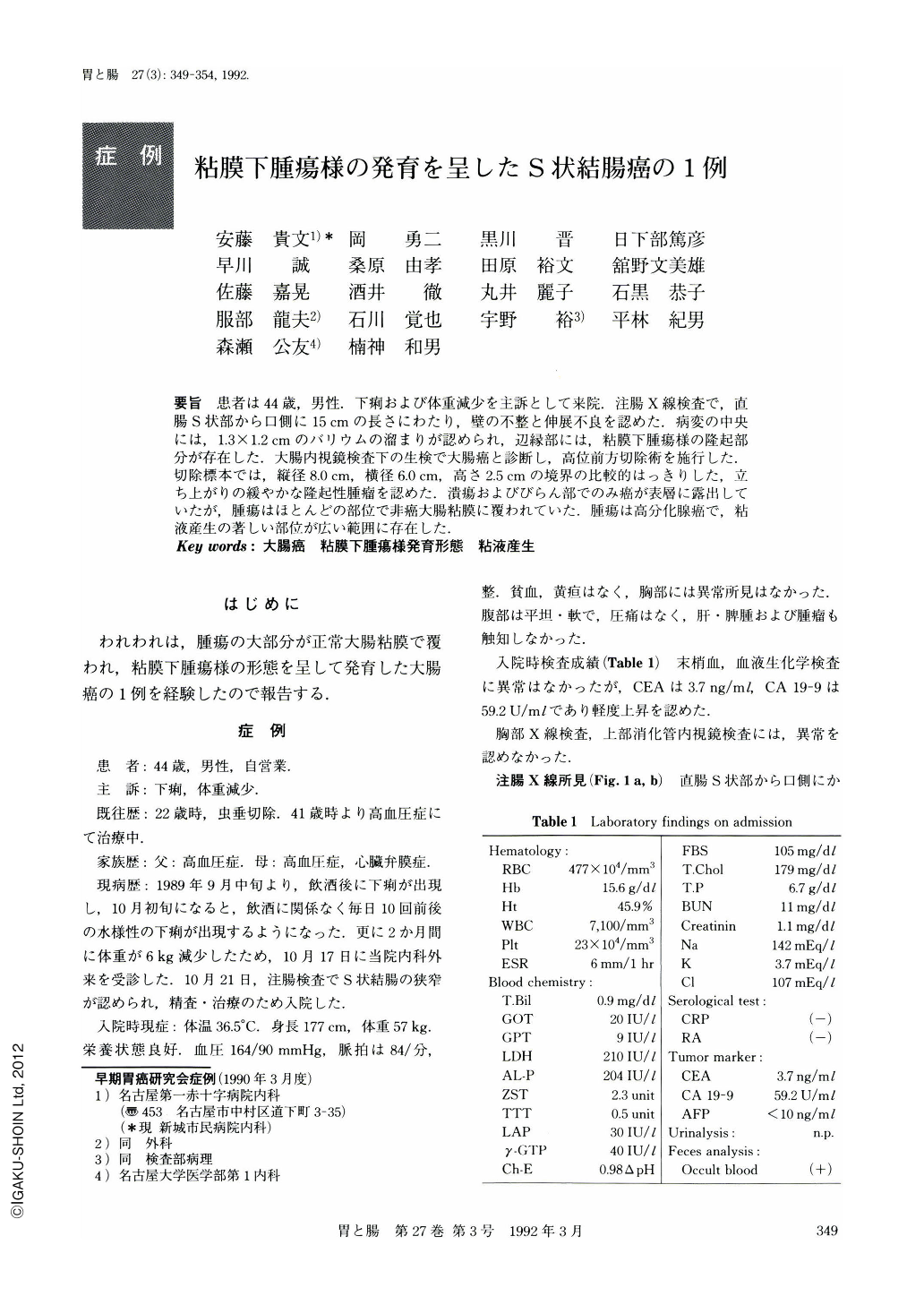
要旨 患者は44歳,男性.下痢および体重減少を主訴として来院.注腸X線検査で,直腸S状部から口側に15cmの長さにわたり,壁の不整と伸展不良を認めた.病変の中央には,1.3×1.2cmのバリウムの溜まりが認められ,辺縁部には,粘膜下腫瘍様の隆起部分が存在した.大腸内視鏡検査下の生検で大腸癌と診断し,高位前方切除術を施行した.切除標本では,縦径8.0cm,横径6.0cm,高さ2.5cmの境界の比較的はっきりした,立ち上がりの緩やかな隆起性腫瘤を認めた.潰瘍およびびらん部でのみ癌が表層に露出していたが,腫瘍はほとんどの部位で非癌大腸粘膜に覆われていた.腫瘍は高分化腺癌で,粘液産生の著しい部位が広い範囲に存在した.
A 44-year-old male patient was admitted to our hospital with the complaints of diarrhea and weight loss. The barium enema examination demonstrated an irregular narrowing with poor distensibility over the length of 15 cm in the distal sigmoid colon. Near in the center of the narrowed segment, there was an ulceration, 1.3 by 1.2 cm in size, surrounded by a wide ulcer bank characteristic of submucosal elevation (Fig. 1). The colonoscopic examination revealed edematous swelling of the mucosa with discrete erythematous and erosive changes in the narrowed segment of the distal sigmoid colon (Fig. 2). Biopsies from the erosions demonstrated adenocarcinoma cells. The resected specimen showed a well defined elevated lesion, 8.0 cm in length, 6.0 cm in width, and 2.5 cm in height, in the distal sigmoid colon, with an ulceration in the center of the lesion (Fig. 3). The margin of the lesion was sharply demarcated but its slope was relatively dull. Most part of the lesion was covered with normal colonic mucosa (Figs. 4 and 7). Histologically, well differentiated adenocarcinoma cells constituted the tumor. Superficially, however, they were limited to the sites of ulceration and erosions in the elevated lesion (Fig. 5). Some mucus-producing cells were present as a component of the tumor cells (Figs. 6b and 7b).
Copyright © 1992, Igaku-Shoin Ltd. All rights reserved.