Japanese
English
- 有料閲覧
- Abstract 文献概要
- 1ページ目 Look Inside
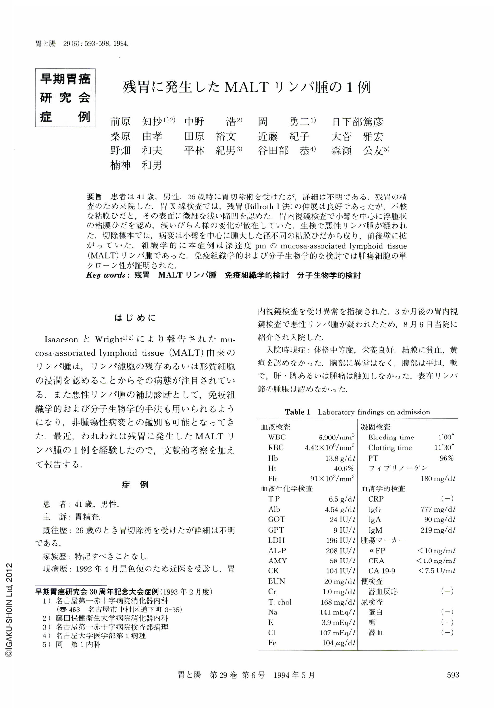
要旨 患者は41歳,男性.26歳時に胃切除術を受けたが,詳細は不明である.残胃の精査のため来院した.胃X線検査では,残胃(Billroth Ⅰ法)の伸展は良好であったが,不整な粘膜ひだと,その表面に微細な浅い陥凹を認めた.胃内視鏡検査で小彎を中心に浮腫状の粘膜ひだを認め,浅いびらん様の変化が散在していた.生検で悪性リンパ腫が疑われた.切除標本では,病変は小彎を中心に腫大した径不同の粘膜ひだから成り,前後壁に拡がっていた.組織学的に本症例は深達度pmのmucosa-associated lymphoid tissue(MALT)リンパ腫であった.免疫組織学的および分子生物学的な検討では腫瘍細胞の単クローン性が証明された.
A 41-year-old man was admitted to our hospital for more detailed evaluation of the stomach. He had an episode of tarry stool four month before admission. He had a history of gastrectomy at the age of 26. The x-ray examination showed a remnant stomach with good extensibility, however, swollen mucosal folds with superficial erosions were observed on the lesser curvature. The endoscopic examination showed irregular mucosal folds with tiny depressions on the lesser curvature. The margin of the lesion was unclear and its center was composed of irregular folds with multiple erosions. The resected stomach showed swollen mucosal folds with erythema and superficial erosions on the lesser curvature. The pathological diagnosis was mucosa associated lymphoid tissue (MALT) lymphoma of intermediate size, diffusely invading the muscularis propria (pm) in the center of the tumor, and the formation of lymphoepithelial lesion was observed in some parts. Immunocytochemical study revealed that most of the tumor cells were positive for CD29, CD20, and λ chain of the surface immunoglobulin. Southern blot analysis demonstrated the clonal rearrangement of the joining region of immunoglobulin heavy chain.

Copyright © 1994, Igaku-Shoin Ltd. All rights reserved.


