Japanese
English
- 有料閲覧
- Abstract 文献概要
- 1ページ目 Look Inside
- サイト内被引用 Cited by
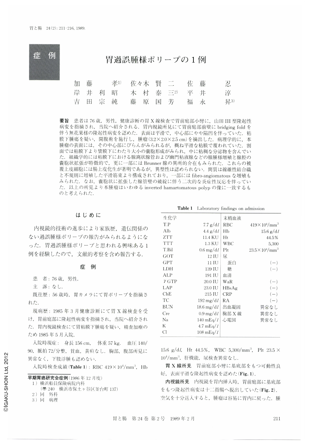
要旨 患者は76歳,男性,健康診断の胃X線検査で胃前庭部小彎に,山田Ⅲ型隆起性病変を指摘され,当院へ紹介される.胃内視鏡所見にて胃前庭部前壁にbridging foldを伴う無花果様の隆起性病変を認めた.表面は平滑で,中心部にやや陥凹を伴っていた.粘膜下腫瘍を疑い,開腹術を施行し,腫瘤(3.2×2.0×2.5cm)を摘出した.病理学的に,本腫瘤の表面には,その中心部にびらんがみられるが,概ね平滑な粘膜で覆われていた.割面では粘膜下より漿膜下にわたり大小の囊胞形成がみられ,中に粘稠な分泌物を含んでいた.組織学的には粘膜下における腺窩状腺管および幽門粘液腺などの腺腫様増殖と腺腔の囊胞状拡張が特徴的で,更に一部にはBrunner腺の異所的介在もみられた.これらの被覆上皮細胞には腸上皮化生が著明であるが,異型性は認められない.間質は線維性結合織と不規則に増殖した平滑筋束より構成されており,一部にはfibro-angiomatousな増殖もみられた.なお,囊胞状に拡張した腺管壁の破綻に伴う二次的な炎症性反応を伴っていた。以上の所見より本腫瘤はいわゆるinverted hamartomatous polypの像に一致するものと考えられた.
A 76-year-old male underwent a medical check up in March of 1985. Upper gastrointestinal x-ray series demonstrated a filling defect at the gastric antrum (Fig. 1). Endoscopic examination revealed Yamada type Ⅲ, fig-shaped polypoid lesion with a smooth surface, prolapsing into the duodenum (Figs. 2 and 3). It was, however easily reduced into the stomach, including the base of the tumor located at the anterior wall of the antrum. Submucosal tumor was suspected because of the presence of bridging fold. He was admitted to the Yokohama Seamen's Insurance Hospital in Map of 1985 with no symptom. Family history was unremarkable for gastrointestinal or pigmentary disorder. Physical examination and laboratory data on admission revealed no abnormality. Surgical resection was carried out. Resected specimen was a soft polypoid tumor, measuring 3.2×2.0×2.5 cm in dimension. It was entirely covered by smooth gastric mucosa except for a central dimple with focal erosion (Fig. 4). Cut section showed a spongy tissue with multiple and various-sized cysts containing mucoid material, which had replaced normal submucosal structure (Fig. 5).
Histologically, the lesion was characterized by an adenomatous proliferation of foveolar glands with or without cystic dilatation (Fig. 6). The lining consists of cuboidal columnar cells with round or oval nuclei located basally and marked intestinal metaplasia but no atypia (Fig. 7a). Between these were foci of hyperplastic pyloric glands and a small islet of Brunner's glands.
The stroma consisted of loose and/or dense fibrous connective tissue and smooth muscle bundles. An angiofibromatous area was also noted (Fig. 8b). Rupture of a large cyst caused a reaction of acute to chronic inflammation.
These histological findings were considered compatible with inverted hamartomatous polyp.

Copyright © 1989, Igaku-Shoin Ltd. All rights reserved.


