Japanese
English
- 有料閲覧
- Abstract 文献概要
- 1ページ目 Look Inside
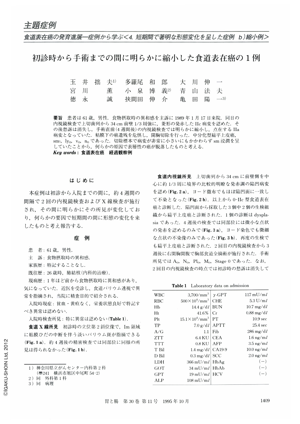
要旨 患者は61歳,男性.食物摂取時の異和感を主訴に1989年1月17日来院.同日の内視鏡検査で上切歯列から34cm前壁1/3周強に,菱形の発赤したⅡc病変を認めた.その後愁訴は消失し,手術直前(4週間後)の内視鏡検査では明らかに縮小し,点在するⅡa病変となっていた,粘膜下の癌遺残を危惧し,開胸切除を行った.中分化型扁平上皮癌,sm1,ly0,V0,n0,であった.切除標本で病変が非常に小さいにもかかわらずsm浸潤を呈していたことから,何らかの原因で表層性の癌が脱落したものと考える.
A 61-year-old man was pointed out to have a depressed lesion in the middle of the esophagus by roentgenographic and endoscopic examinations. He complained of post-sternal discomfort during eating. The lesion had reddened, and occupied one-third of the circumference of the esophagus. This lesion was diagnosed as moderately differentiated squamous cell carcinoma by biopsy.
Four weeks later the lesion had apparently reduced in size, and was detected by iodine staining only with difficulty. In the resected specimen there were four small elevated lesions, measuring 0.2, 0.2, 0.2 and 0.4 cm in size respectively.
What reduced the lesion? It was suspected that the iodine spraying and the taking of biopsy specimens etc. may have caused the reduction, but we also thought that it could be attributed to natural courses.

Copyright © 1995, Igaku-Shoin Ltd. All rights reserved.


