Japanese
English
- 有料閲覧
- Abstract 文献概要
- 1ページ目 Look Inside
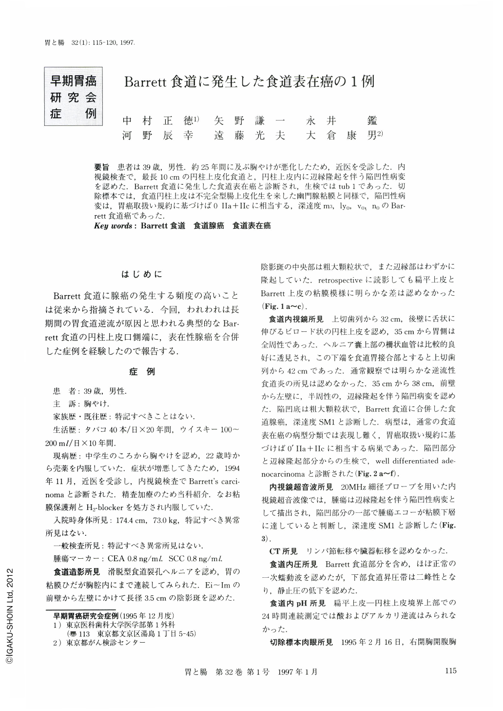
要旨 患者は39歳,男性.約25年間に及ぶ胸やけが悪化したため,近医を受診した.内視鏡検査で,最長10cmの円柱上皮化食道と,円柱上皮内に辺縁隆起を伴う陥凹性病変を認めた.Barrett食道に発生した食道表在癌と診断され,生検ではtub 1であった.切除標本では,食道円柱上皮は不完全型腸上皮化生を来した幽門腺粘膜と同様で,陥凹性病変は,胃癌取扱い規約に基づけば0 Ⅱa+Ⅱcに相当する,深達度m3,ly0,v0,n0のBarrett食道癌であった.
A 39-year-old man visited our hospital with a longstanding history of heartburn. Endoscopic examination revealed a columnar epithelium-lined esophagus for 10 cm at its greatest length and a depressed lesion surrounded by an elevated component. The biopsy specimen from the lesion was diagnosed as well-differentiated adenocarcinoma. The subtotal esophagectomy through right thoractomy was performed.
Histological studies of the resected specimen demonstrated that the columnar epithelium had intestinal metaplasia and the tumorous lesion was Barrett's carcinoma limited to the mucosa (m3), expressed 0-Ⅱa + Ⅱc type based on the classification of early gastric carcinoma. Neither vascular spreading nor lymph node involvement was identified.

Copyright © 1997, Igaku-Shoin Ltd. All rights reserved.


