Japanese
English
- 有料閲覧
- Abstract 文献概要
- 1ページ目 Look Inside
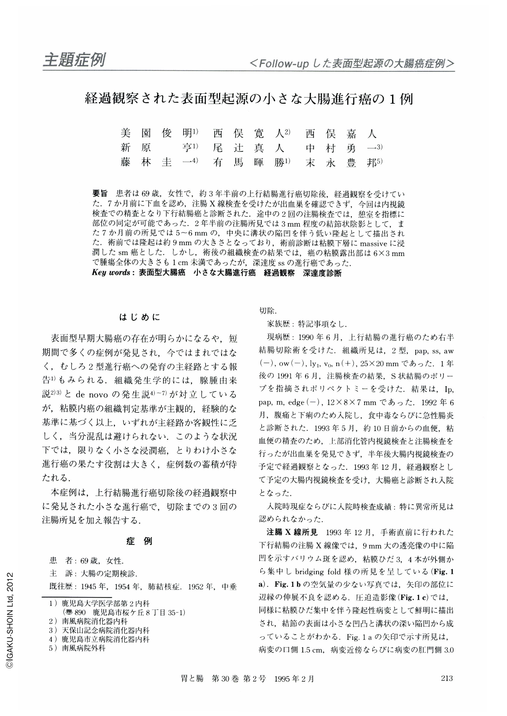
要旨 患者は69歳,女性で,約3年半前の上行結腸進行癌切除後,経過観察を受けていた.7か月前に下血を認め,注腸X線検査を受けたが出血巣を確認できず,今回は内視鏡検査での精査となり下行結腸癌と診断された.途中の2回の注腸検査では,憩室を指標に部位の同定が可能であった.2年半前の注腸所見では3mm程度の結節状陰影として,また7か月前の所見では5~6mmの,中央に溝状の陥凹を伴う低い隆起として描出された.術前では隆起は約9mmの大きさとなっており,術前診断は粘膜下層にmassiveに浸潤したsm癌とした.しかし,術後の組織検査の結果では,癌の粘膜露出部は6×3mmで腫瘍全体の大きさもlcm未満であったが,深達度ssの進行癌であった.
A 69-year-old woman, who underwent partial colectomy about 3 years before to resect an advanced cancer of the ascending colon, visited our hospital for follow-up examination of the colon. A flat elevated lesion with central depression was detected in the descending colon by colonoscopy and barium enema study. Preoperative diagnosis was an early cancer of type Ⅱa + Ⅱc with massive invasion but limited to the submucosa.
Partial colectomy was performed again, and histological examination showed that the lesion was tubular adenocarcinoma without adenomatous components, deeply infiltrating the subserosa. The size of the cancer exposed to the colonic lumen measured approximately 6 × 3 mm.
In the period between these operations, barium enema studies were performed twice. Seven months prior to this surgery, the patient had complained of an episode of bloody stool. Though it was overlooked at the time, a small round flat elevated shadow with central depression was clearly demonstrated on the x-ray pictures. The lesion on the x-ray pictures was about half the size of the lesion found in the final study.
Moreover, a careful retrospective study enabled us to go back and see the same lesion as a small nodular shadow about 3 mm in diameter on the x-ray pictures taken 2.5 years before the final operation. These findings of follow-up barium enema studies suggest that this cancer developed rapidly in the late stage, but only slowly in the early stage.
Detection of any small flat elevated lesion less than 5 mm in diameter can be regarded as a basis for early diagnosis of colon cancer.

Copyright © 1995, Igaku-Shoin Ltd. All rights reserved.


