Japanese
English
- 有料閲覧
- Abstract 文献概要
- 1ページ目 Look Inside
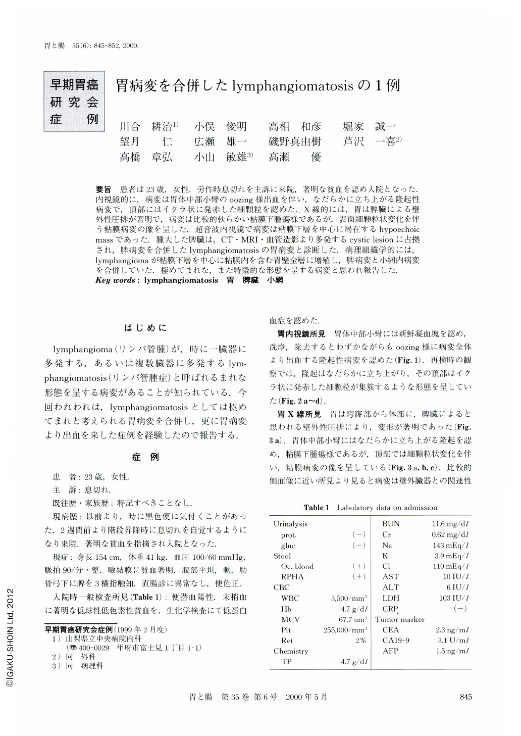
要旨 患者は23歳,女性.労作時息切れを主訴に来院,著明な貧血を認め入院となった.内視鏡的に,病変は胃体中部小彎のoozing様出血を伴い,なだらかに立ち上がる隆起性病変で,頂部にはイクラ状に発赤した細顆粒を認めた.X線的には,胃は脾臓による壁外性圧排が著明で,病変は比較的軟らかい粘膜下腫瘍様であるが,表面細顆粒状変化を伴う粘膜病変の像を呈した.超音波内視鏡で病変は粘膜下層を中心に局在するhypoechoicmassであった.腫大した脾臓は,CT・MRI・血管造影より多発するcystic lesionに占拠され,脾病変を合併したlymphangiomatosisの胃病変と診断した.病理組織学的には,lymphangiomaが粘膜下層を中心に粘膜内を含む胃壁全層に増殖し,脾病変と小網内病変を合併していた.極めてまれな,また特徴的な形態を呈する病変と思われ報告した.
A 23-year-old female was admitted because of exertional dyspnea and severe anemia. Endoscopic examination disclosed a protruding lesion covered with a gathering of smooth and reddish granules, on the lesser curvature of the gastric body. It also disclosed hemorrhage or oozing from this lesion. X-ray examination revealed a flat and smooth protrusion covered with a gathering of granules and it appeared to be soft because the change of shape seen in different photos. The stomach was poorly outlined with marked extrinsic compression by the enlarged spleen. Endoscopic ultrasonography revealed the lesion as a relatively hypoechoic and heterogeneous mass mainly occupying the submucosal layer. Abdominal dynamic CT scan with administration of contrast medium showed multiple cystic areas occupying the enlarged spleen with low vascular density. MRI shows multiple mass lesions with low intensity by T1-and high intensity by T2-weighted images. In addition, it was enhanced by administration of gadolinium on T1-weighted image. Splenic angiography demonstrated a marked stretching of intrasplenic vessels in the arterial phase and “Swiss cheese” appearance in the tissue phase.
We diagnosed the above lesions as the result of both gastric and splenic lymphangiomatosis, and the patient underwent partial gastrectomy combined with splenectomy. Macro- and microscopic examinations after surgery revealed that the lymphangiomatosis had affected the stomach, spleen and lesser omentum. Microscopically, the gastric lesion showed transmucosal proliferation of lymphangioma containing amounts of capillaries in their stroma, and which were causative of the hemorrhage. The gastric lesion of lymphangiomatosis has not been reported in the literature in English yet, and this is only the second report in Japanese medical literattre

Copyright © 2000, Igaku-Shoin Ltd. All rights reserved.


