Japanese
English
- 有料閲覧
- Abstract 文献概要
- 1ページ目 Look Inside
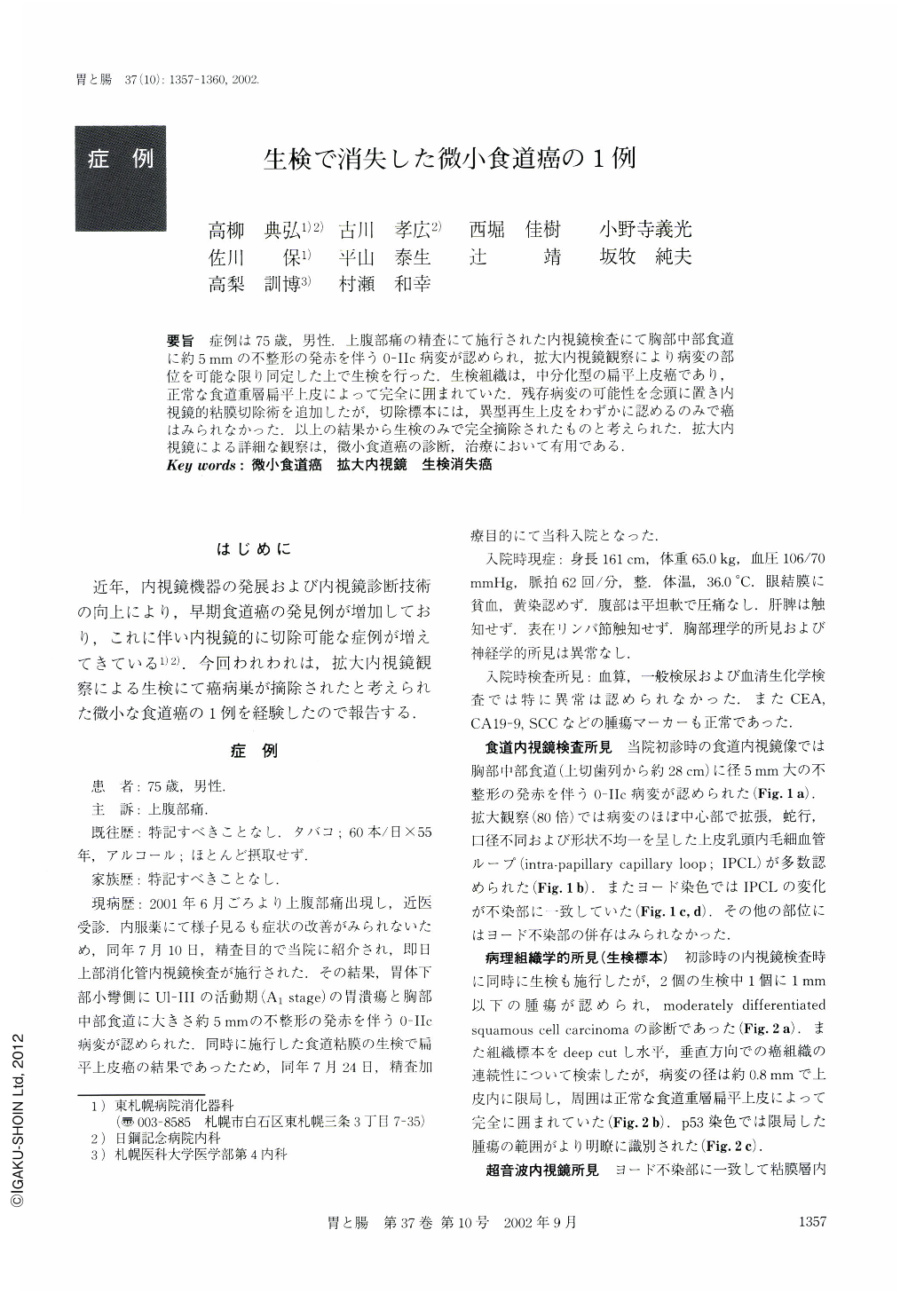
要旨 症例は75歳,男性.上腹部痛の精査にて施行された内視鏡検査にて胸部中部食道に約5mmの不整形の発赤を伴う0-Ⅱc病変が認められ,拡大内視鏡観察により病変の部位を可能な限り同定した上で生検を行った.生検組織は,中分化型の扁平上皮癌であり,正常な食道重層扁平上皮によって完全に囲まれていた.残存病変の可能性を念頭に置き内視鏡的粘膜切除術を追加したが,切除標本には,異型再生上皮をわずかに認めるのみで癌はみられなかった.以上の結果から生検のみで完全摘除されたものと考えられた.拡大内視鏡による詳細な観察は,微小食道癌の診断,治療において有用である.
We have recently encountered a minute esophageal cancer which disappeared due to biopsy. A 75-year-old man was referred to our hospital for examination of epigastralgia. Endoscopic examination revealed a small irregular red-spot in the middle esophagus where one of two biopsied specimens obtained led to a diagnosis of moderately differentiated squamous cell carcinoma. The affected region was treated by endoscopic mucosal resection. In resected specimens, only a small focus of atypical epithelium was noted, and no cancerous focus was found despite thorough pathological examination. It was suggested that high magnification endoscopic examination was useful in detecting such a minute carcinoma of the esophagus.

Copyright © 2002, Igaku-Shoin Ltd. All rights reserved.


