Japanese
English
- 有料閲覧
- Abstract 文献概要
- 1ページ目 Look Inside
- サイト内被引用 Cited by
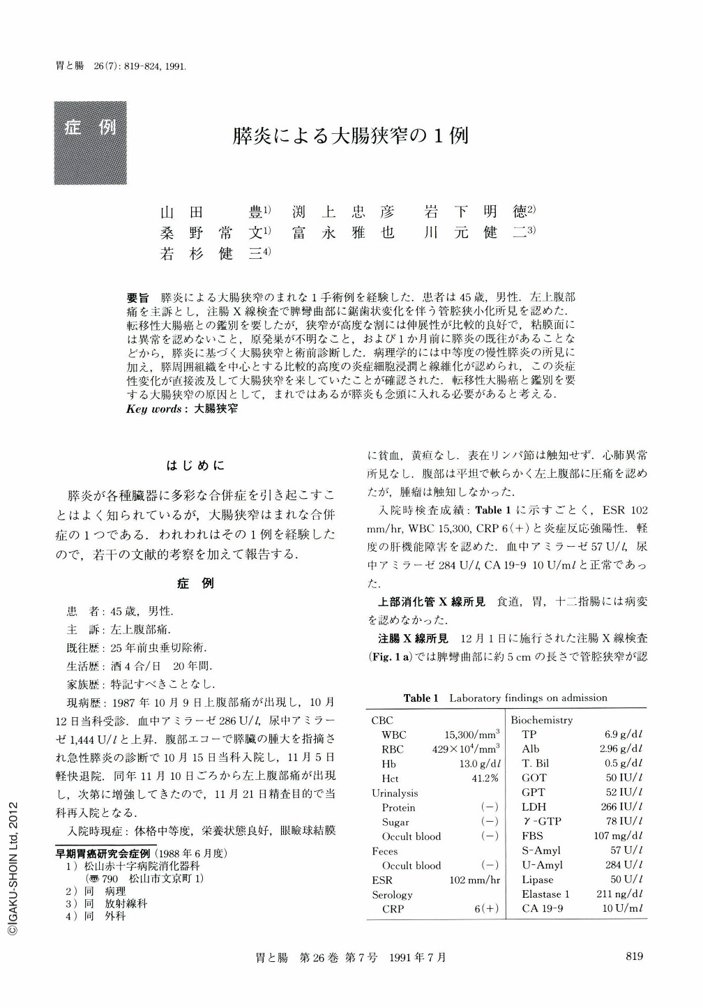
要旨 膵炎による大腸狭窄のまれな1手術例を経験した.患者は45歳,男性.左上腹痛を主訴とし,注腸X線検査で脾彎曲部に鋸歯状変化を伴う管腔狭小化所見を認めた.転移性大腸癌との鑑別を要したが,狭窄が高度な割には伸展性が比較的良好で,粘膜面には異常を認めないこと,原発巣が不明なこと,および1か月前に膵炎の既往があることなどから,膵炎に基づく大腸狭窄と術前診断した.病理学的には中等度の慢性膵炎の所見に加え,膵周囲組織を中心とする比較的高度の炎症細胞浸潤と線維化が認められ,この炎症性変化が直接波及して大腸狭窄を来していたことが確認された.転移性大腸癌と鑑別を要する大腸狭窄の原因として,まれではあるが膵炎も念頭に入れる必要があると考える.
We presented a case of colonic stenosis associated with pancreatitis. A 45-year-old man visited our hospital complaining of left upper abdominal pain. Barium enema study revealed a narrowing with marginal serration in the splenic flexure (Fig. 1a). The distensibility of the lumen was relatively good and the mucosal surface was intact (Fig. 1b). Endoscopically, the mucosa was edematous and ulceration was not seen (Fig. 3a). The lumen was distended by infusion of air (Fig. 3b). No signs suggestive of malignancy were found either by abdominal CT scan, ERCP or angiogram. He had a past history of pancreatitis one month prior to the admission. Thus, we considered the lesion as inflammatory preoperatively. Barium enema performed 18 days later from the first examination revealed an obstruction (Fig. 2) and operation was performed. Histologically, mild chronic inflammatory infiltrates and moderate fibrosis were shown in the pancreatic tissue (Fig. 9). Similar inflammatory change and moderate to marked fibrosis were also shown in the colonic serosa (Fig. 10). It was suggested that the colonic stenosis associated with pancreatitis developed from pericolitis.

Copyright © 1991, Igaku-Shoin Ltd. All rights reserved.


