Japanese
English
- 有料閲覧
- Abstract 文献概要
- 1ページ目 Look Inside
- サイト内被引用 Cited by
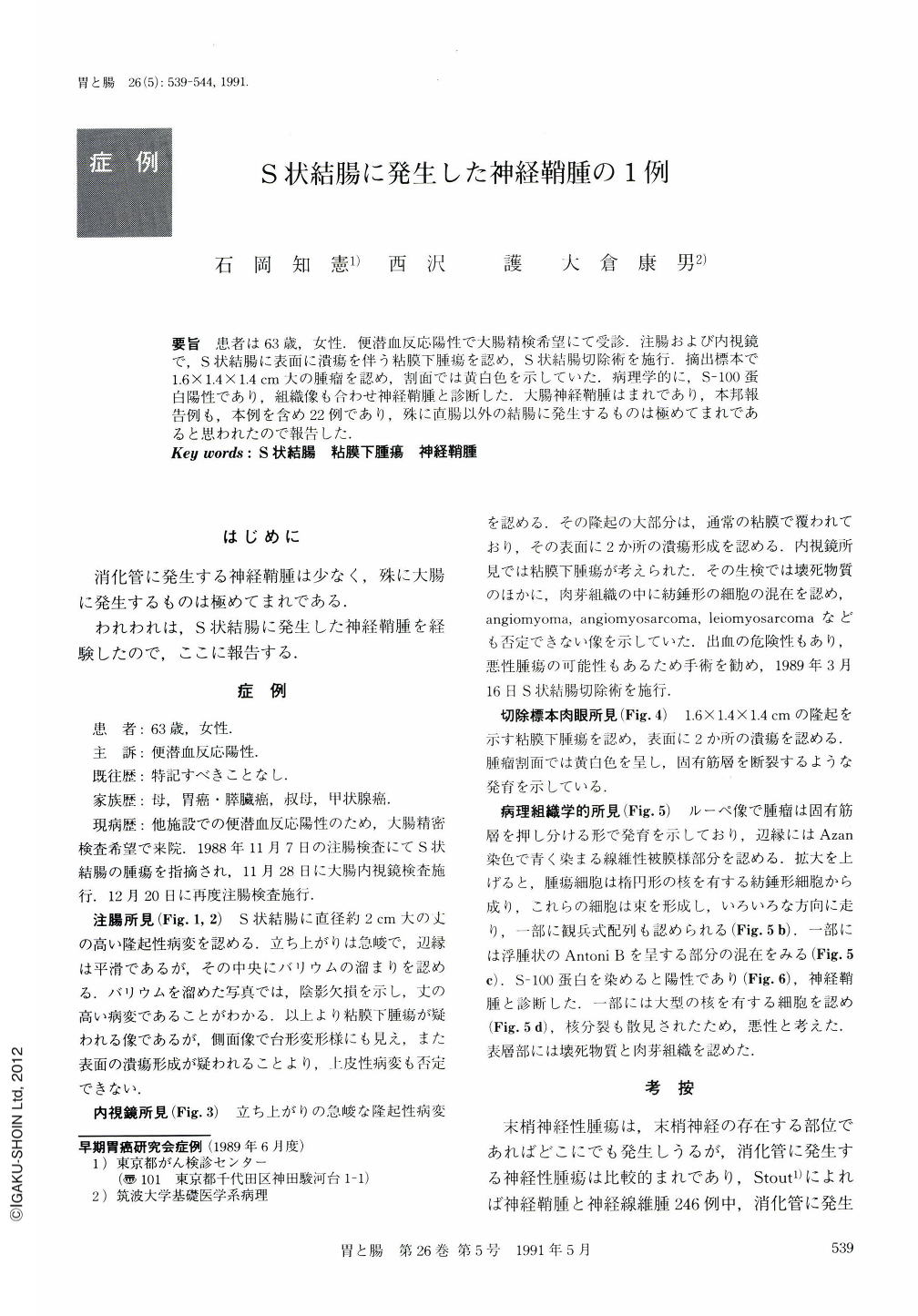
要旨 患者は63歳,女性.便潜血反応陽性で大腸精検希望にて受診.注腸および内視鏡で,S状結腸に表面に潰瘍を伴う粘膜下腫瘍を認め,S状結腸切除術を施行.摘出標本で1.6×1.4×1.4cm大の腫瘤を認め,割面では黄白色を示していた.病理学的に,S-100蛋白陽性であり,組織像も合わせ神経鞘腫と診断した.大腸神経鞘腫はまれであり,本邦報告例も,本例を含め22例であり,殊に直腸以外の結腸に発生するものは極めてまれであると思われたので報告した.
A 63-year-old female underwent barium enema and colonofiberscopy because of positive fecal occult blood. An apparently elevated lesion with a central depression was visualized in the sigmoid colon by barium enema (Figs. 1 and 2). Profile appearance showed a gently rising elevation indicative of submucosal tumor. The margin was rigid at the bottom of the lesion, suggesting that epithelial lesion could not be entirely ruled out. The lesion was diagnosed as submucosal tumor by endoscopy based on a variety of its characteristics (Fig. 3). Sigmoid colectomy was performed. In the resected specimen was a submucosal tumor with a central depressions, measuring 1.6×1.4×1.4 cm in size (Fig. 4). The cut surface of the tumor had yellowish hue, which was considered to be one of the most crucial characteristics of schwannoma. Histological examination showed interlacing fascicular pattern (Fig. 5 a) and palisading of nuclei (Fig. 5 b). Loose, reticular and poor cellular patterns corresponded to Antoni type B (Fig. 5 c). In addition, the tumor was positive for S-100 protein staining (Fig. 6). Based on these findings the lesion was diagnosed as schwannoma.
Only 21 cases of schwannoma of the colo-rectum have been reported to date in Japan, 14 of which arising from the rectum (Table 1). Schwannoma of the colon as we reported here is thus rare.

Copyright © 1991, Igaku-Shoin Ltd. All rights reserved.


