Japanese
English
- 有料閲覧
PPV購入案内
1,320円 (1,200円+税10%) こちらは、349ページから354ページの文献です。購入には医書.jp本会員登録が必要です。
会員登録完了後に改めて上記「購入サイトへ」を選択してください。
または「購入サイトへ」選択後に「購入ログイン」、「新規会員登録」の順に進んでください。
会員登録について詳しくはをご確認ください。
- Abstract 文献概要
- 1ページ目 Look Inside
- 参考文献 Reference
- サイト内被引用 Cited by
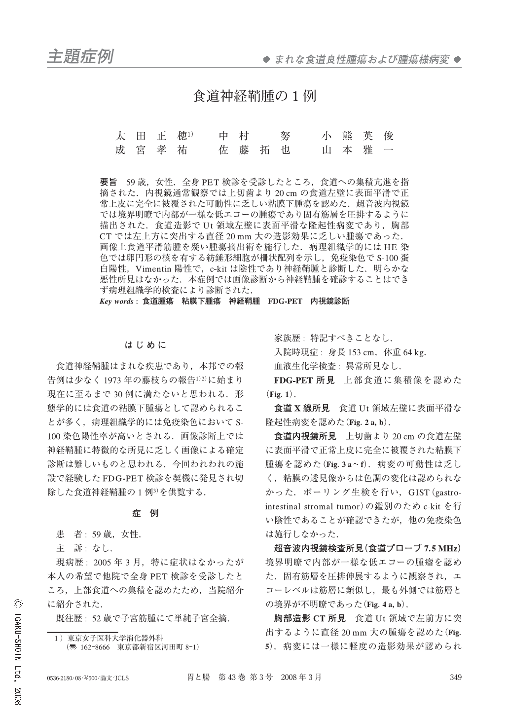
要旨 59歳,女性.全身PET検診を受診したところ,食道への集積亢進を指摘された.内視鏡通常観察では上切歯より20cmの食道左壁に表面平滑で正常上皮に完全に被覆された可動性に乏しい粘膜下腫瘍を認めた.超音波内視鏡では境界明瞭で内部が一様な低エコーの腫瘍であり固有筋層を圧排するように描出された.食道造影でUt領域左壁に表面平滑な隆起性病変であり,胸部CTでは左上方に突出する直径20mm大の造影効果に乏しい腫瘍であった.画像上食道平滑筋腫を疑い腫瘍摘出術を施行した.病理組織学的にはHE染色では卵円形の核を有する紡錘形細胞が柵状配列を示し,免疫染色でS-100蛋白陽性,Vimentin陽性で,c-kitは陰性であり神経鞘腫と診断した.明らかな悪性所見はなかった.本症例では画像診断から神経鞘腫を確診することはできず病理組織学的検査により診断された.
A 59-year-old woman underwent medical screening and FDG-PET detected uptake by a lesion in the thoracic esophagus. Endoscopy showed a smooth submucosal tumor that was completely covered with normal mucosa, while endoscopic ultrasonography revealed a well-demarcated and uniformly low echoic tumor compressing the muscle layer. Barium meal showed a smooth elevated lesion in the upper thoracic esophagus, while CT revealed a tumor 20 mm in diameter that projected to the left of the esophageal wall and was poorly enhanced. Leiomyoma was suspected, so enucleation was performed. Histopathological examination showed spindle-shaped cells with a palisading arrangement, while immunohistochemistry demonstrated positive staining for S-100 protein and vimentin. Accordingly, the pathological diagnosis of the tumor was schwannoma.
Copyright © 2008, Igaku-Shoin Ltd. All rights reserved.