Japanese
English
- 有料閲覧
- Abstract 文献概要
- 1ページ目 Look Inside
- 参考文献 Reference
- サイト内被引用 Cited by
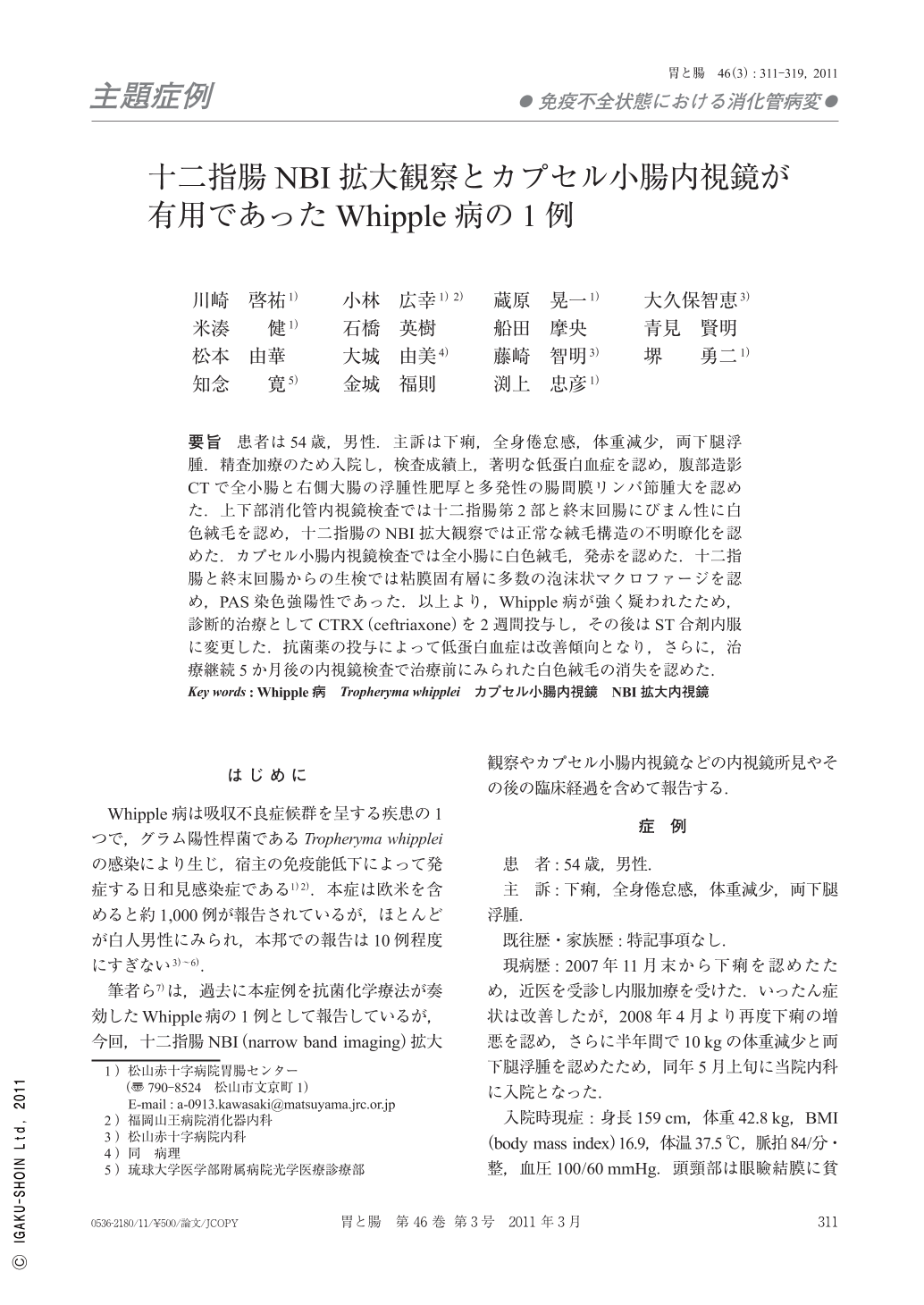
要旨 患者は54歳,男性.主訴は下痢,全身倦怠感,体重減少,両下腿浮腫.精査加療のため入院し,検査成績上,著明な低蛋白血症を認め,腹部造影CTで全小腸と右側大腸の浮腫性肥厚と多発性の腸間膜リンパ節腫大を認めた.上下部消化管内視鏡検査では十二指腸第2部と終末回腸にびまん性に白色絨毛を認め,十二指腸のNBI拡大観察では正常な絨毛構造の不明瞭化を認めた.カプセル小腸内視鏡検査では全小腸に白色絨毛,発赤を認めた.十二指腸と終末回腸からの生検では粘膜固有層に多数の泡沫状マクロファージを認め,PAS染色強陽性であった.以上より,Whipple病が強く疑われたため,診断的治療としてCTRX(ceftriaxone)を2週間投与し,その後はST合剤内服に変更した.抗菌薬の投与によって低蛋白血症は改善傾向となり,さらに,治療継続5か月後の内視鏡検査で治療前にみられた白色絨毛の消失を認めた.
A 54-year-old man was admitted to our institution because of diarrhea, general fatigue, weight loss, and lower edema in both things. Computed tomography of the abdomen revealed thickening of the small-bowel wall and enlarged mesenteric lymph nodes. Endoscopy disclosed diffuse yellow-white shaggy mucosa of the duodenum, jejunum, and ileum. The villi of the duodenal mucosa were invisible on NBI(narrow band imaging)magnifying observation. On histological examination of the biopsy specimens, the lamina propria of the intestinal mucosa was seemed to be heavily infiltrated by foamy macrophages that were PAS(periodic acid-Schiff)-positive. Based on characteristic pathological findings, we thought that the clinical diagnosis could be Whipple's disease. The patient was treated with a two-week course of CTRX(ceftriaxone), followed by trimethoprim-sulfamethoxazole. After 24-days of therapy, clinical symptoms and hypoproteinemia had improved. At 5-month follow up, diffuse yellow-white shaggy mucosa had improved dramatically and villi of the duodenal mucosa were visible on endoscopic examination.
Copyright © 2011, Igaku-Shoin Ltd. All rights reserved.