Japanese
English
- 有料閲覧
- Abstract 文献概要
- 1ページ目 Look Inside
- 参考文献 Reference
- サイト内被引用 Cited by
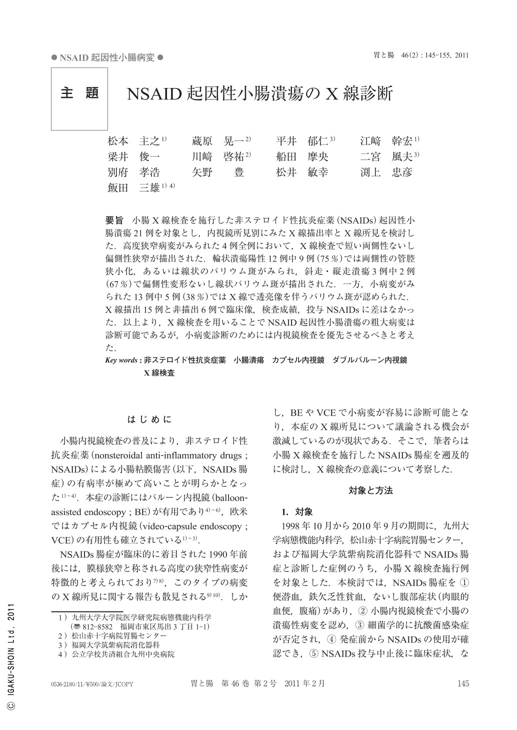
要旨 小腸X線検査を施行した非ステロイド性抗炎症薬(NSAIDs)起因性小腸潰瘍21例を対象とし,内視鏡所見別にみたX線描出率とX線所見を検討した.高度狭窄病変がみられた4例全例において,X線検査で短い両側性ないし偏側性狭窄が描出された.輪状潰瘍陽性12例中9例(75%)では両側性の管腔狭小化,あるいは線状のバリウム斑がみられ,斜走・縦走潰瘍3例中2例(67%)で偏側性変形ないし線状バリウム斑が描出された.一方,小病変がみられた13例中5例(38%)ではX線で透亮像を伴うバリウム斑が認められた.X線描出15例と非描出6例で臨床像,検査成績,投与NSAIDsに差はなかった.以上より,X線検査を用いることでNSAID起因性小腸潰瘍の粗大病変は診断可能であるが,小病変診断のためには内視鏡検査を優先させるべきと考えた.
We retrospectively investigated radiographic and enteroscopic findings in 21 patients with nonsteroidal anti-inflammatory drugs-induced enteropathy(NSAIDs-enteropathy). Endoscopically, there were 4 cases of diaphragm-like stricture, 12 cases of circular ulcers, 3 cases of oblique ulcers, and 13 cases of diminutive ulcers. Radiography depicted diaphragm-like stricture as severe and concentric stenosis in all the 6 cases. Circular ulcers were depicted as tubular narrowings in 9 cases(75%)and oblique ulcers were depicted as eccentric deformity or linear barium flecks in 2 cases(67%). However, radiography could depict diminutive ulcers as small barium flecks in only 5 cases(38%). There was no difference in clinical features between radiographically positive and negative cases. These findings suggest that, even though radiography could depict advanced lesions in the disease, enteroscopy seems to be the first choice of procedure for the diagnosis of NSAIDs-enteropathy.

Copyright © 2011, Igaku-Shoin Ltd. All rights reserved.


