Japanese
English
- 有料閲覧
- Abstract 文献概要
- 1ページ目 Look Inside
- 参考文献 Reference
- サイト内被引用 Cited by
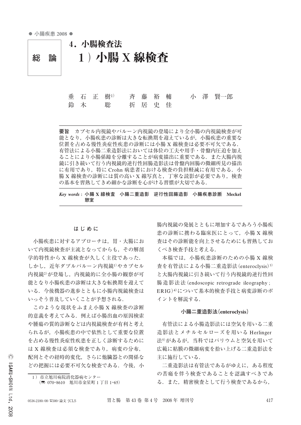
要旨 カプセル内視鏡やバルーン内視鏡の登場により全小腸の内視鏡検査が可能となり,小腸疾患の診断は大きな転換期を迎えているが,小腸疾患の重要な位置を占める慢性炎症性疾患の診断には小腸X線検査は必要不可欠である.有管法による小腸二重造影法においては体位の工夫や用手・骨盤内圧迫を加えることにより小腸係蹄を分離することが病変描出に重要である.また大腸内視鏡に引き続いて行う内視鏡的逆行性回腸造影法は骨盤内回腸の微細所見の描出に有用であり,特にCrohn病患者における検査の負担軽減に有用である.小腸X線検査の診断には質の高いX線写真と,丁寧な読影が必要であり,検査の基本を習熟してきめ細かな診断を心がける習慣が大切である.
Development of capsule endoscopy and balloon enteroscopy that enable total enteroscopy has become a turning point in the diagnosis of intestinal disease. However radiographic examination is still essential for the diagnosis of chronic inflammatory diseases of the small intestine, which gives it an important role in the diagnosis of small intestinal diseases. There are very important techniques in order to obtain good images in small bowel enteroclysis, such as changing to adequate positions and additional compression by hand or towel to prevent overlapping loops. Endoscopic retrograde ileography is a useful modality for the delineation of faint abnormalities in the ileum and especially for the reduction of the burden of examination by the combination of colonoscopy and radiographic examination. Skilled technique, and detailed and careful interpretation of data for the precise diagnosis of small intestinal diseases, are essential for high quality radiography.

Copyright © 2008, Igaku-Shoin Ltd. All rights reserved.


