Japanese
English
- 有料閲覧
PPV購入案内
1,320円 (1,200円+税10%) こちらは、1270ページから1282ページの文献です。購入には医書.jp本会員登録が必要です。
会員登録完了後に改めて上記「購入サイトへ」を選択してください。
または「購入サイトへ」選択後に「購入ログイン」、「新規会員登録」の順に進んでください。
会員登録について詳しくはをご確認ください。
- Abstract 文献概要
- 1ページ目 Look Inside
- 参考文献 Reference
- サイト内被引用 Cited by
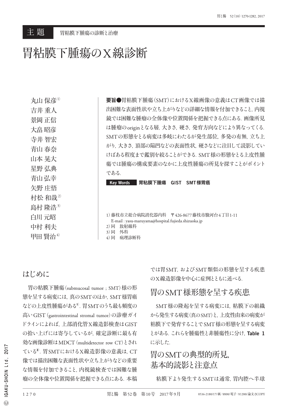
要旨●胃粘膜下腫瘍(SMT)におけるX線画像の意義はCT画像では描出困難な表面性状や立ち上がりなどの詳細な情報を付加できること,内視鏡では困難な腫瘤の全体像や位置関係を把握できる点にある.画像所見は腫瘤のoriginとなる層,大きさ,硬さ,発育方向などにより異なってくる.SMTの形態をとる病変は多岐にわたるが発生部位,多発の有無,立ち上がり,大きさ,頂部の陥凹などの表面性状,硬さなどに注目して読影していけばある程度まで鑑別を絞ることができる.SMT様の形態をとる上皮性腫瘍では腫瘍の構成要素のなかに上皮性腫瘍の所見を探すことがポイントである.
The importance of radiological examination in the diagnosis of gastric SMTs(submucosal tumors)are highly-detailed observation of the mucosal surface pattern and rising shoulder, which are difficult to evaluate using computed tomography examination, along with recognition of the whole overview and positional relationships in the stomach, which is difficult to grasp via endoscopic study. Radiological findings differ depending on the layer of origin, size, the degree of hardness, and the direction of tumor growth. A variety of these lesions show an SMT-like appearance. Differential diagnosis of these lesions requires precise observation of the location, multiplicity, rising shoulder, size, depression, and the hardness of the tumor. Such findings from mucosal tumors are important in the diagnosis of SMT resembling mucosal tumors.
Copyright © 2017, Igaku-Shoin Ltd. All rights reserved.