Japanese
English
- 有料閲覧
- Abstract 文献概要
- 1ページ目 Look Inside
- 参考文献 Reference
- サイト内被引用 Cited by
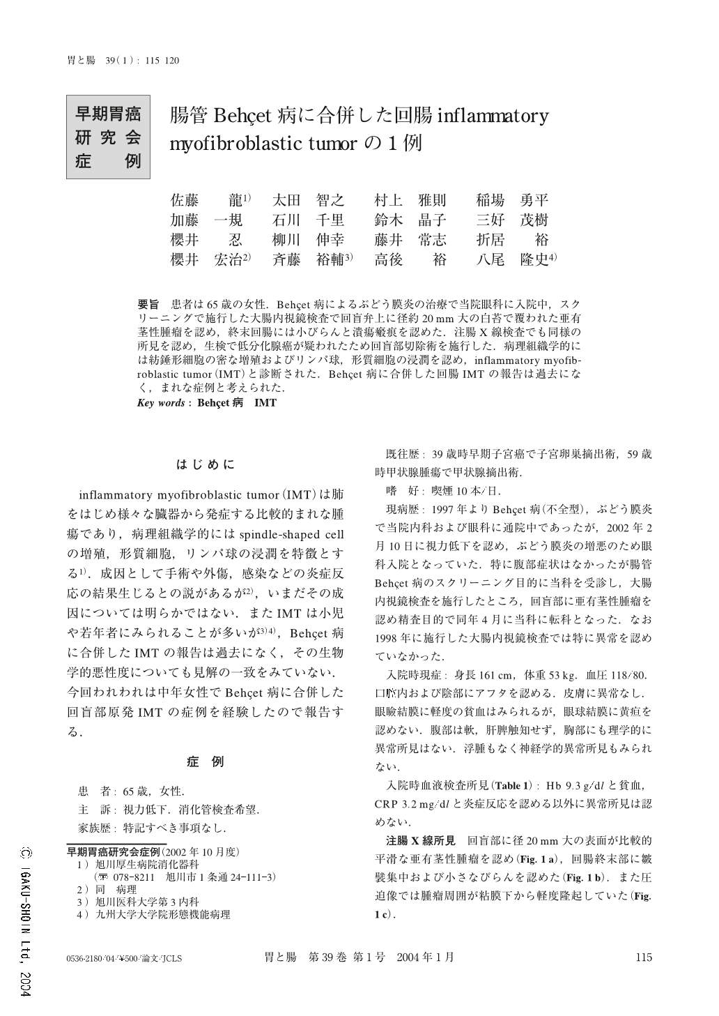
要旨 患者は65歳の女性.Behҫet病によるぶどう膜炎の治療で当院眼科に入院中,スクリーニングで施行した大腸内視鏡検査で回盲弁上に径約20mm大の白苔で覆われた亜有茎性腫瘤を認め,終末回腸には小びらんと潰瘍瘢痕を認めた.注腸X線検査でも同様の所見を認め,生検で低分化腺癌が疑われたため回盲部切除術を施行した.病理組織学的には紡錘形細胞の密な増殖およびリンパ球,形質細胞の浸潤を認め,inflammatory myofibroblastic tumor(IMT)と診断された.Behҫet病に合併した回腸IMTの報告は過去になく,まれな症例と考えられた.
A 65-year-old woman, who had been treated for ocular Behҫet's disease since 1997, visited our unit, without any complaint, for intestinal screening in March, 2002. Colonoscopic examination showed a subpedunculated tumor measuring 20 mm in size covered with white coating on the ileocecal valve. Ulcer scars and small erosions were also seen in the terminal ileum. Barium enema study revealed the same polypoid lesion and inflammation in the ileocecum. Although the tumor was diagnosed as an inflammatory polyp according to the above findings, since it could not be ruled out as a malignant epithelial tumor, ileocecal resection was performed in May, 2002. Histological examination showed that the tumor consisted of spindle-shaped cell proliferation with a scattering of nuclear mitosis, lymphocytes and plasma cell infiltration based on severe submucosal fibrosis. Additionally non-specific erosions or ulcer scars were noted in the terminal ileum. Immunohistochemical study revealed that the tumor was positive for vimentin, and cytokeratin, but negative for ALK. Finally this tumor was diagnosed as IMT with intermediate malignant potential developed from intestinal Behҫet's disease.
1) Department of Gastroenterology, Asahikawa Kosei General Hospital, Asahikawa, Japan

Copyright © 2004, Igaku-Shoin Ltd. All rights reserved.


