Japanese
English
- 有料閲覧
- Abstract 文献概要
- 1ページ目 Look Inside
- 参考文献 Reference
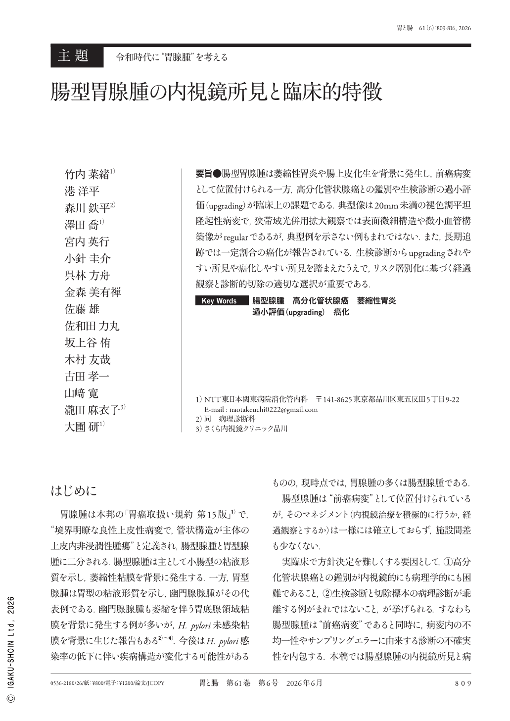
要旨●腸型胃腺腫は萎縮性胃炎や腸上皮化生を背景に発生し,前癌病変として位置付けられる一方,高分化管状腺癌との鑑別や生検診断の過小評価(upgrading)が臨床上の課題である.典型像は20mm未満の褪色調平坦隆起性病変で,狭帯域光併用拡大観察では表面微細構造や微小血管構築像がregularであるが,典型例を示さない例もまれではない.また,長期追跡では一定割合の癌化が報告されている.生検診断からupgradingされやすい所見や癌化しやすい所見を踏まえたうえで,リスク層別化に基づく経過観察と診断的切除の適切な選択が重要である.
Intestinal-type gastric adenoma, a premalignant lesion arising in atrophic gastritis with intestinal metaplasia, poses key challenges in clinical practice. These include the challenge in endoscopic differentiation from well-differentiated gastric adenocarcinoma and histologic underestimation on forceps biopsy, which can lead to diagnostic upgrading after endoscopic resection. Typical endoscopic findings include a pale, flat, slightly elevated lesion measuring less than 20mm. On magnifying endoscopy with narrow-band imaging, lesions generally exhibit regular microsurface and microvascular patterns, with characteristic features such as a white opaque substance and dense-type crypt openings. However, the presence of erythema, depression, nodularity, and irregular microvascular and/or microsurface patterns should raise suspicion for coexisting carcinoma. Long-term follow-up studies indicate that many cases of gastric adenomas are eventually diagnosed as carcinoma during surveillance, highlighting the importance of distinguishing true malignant transformation from initial histologic underestimation. Balancing careful surveillance with diagnostic/therapeutic endoscopic resection based on endoscopic risk features is essential for an individualized management strategy.
Copyright © 2026, Igaku-Shoin Ltd. All rights reserved.