Japanese
English
- 有料閲覧
- Abstract 文献概要
- 1ページ目 Look Inside
- 参考文献 Reference
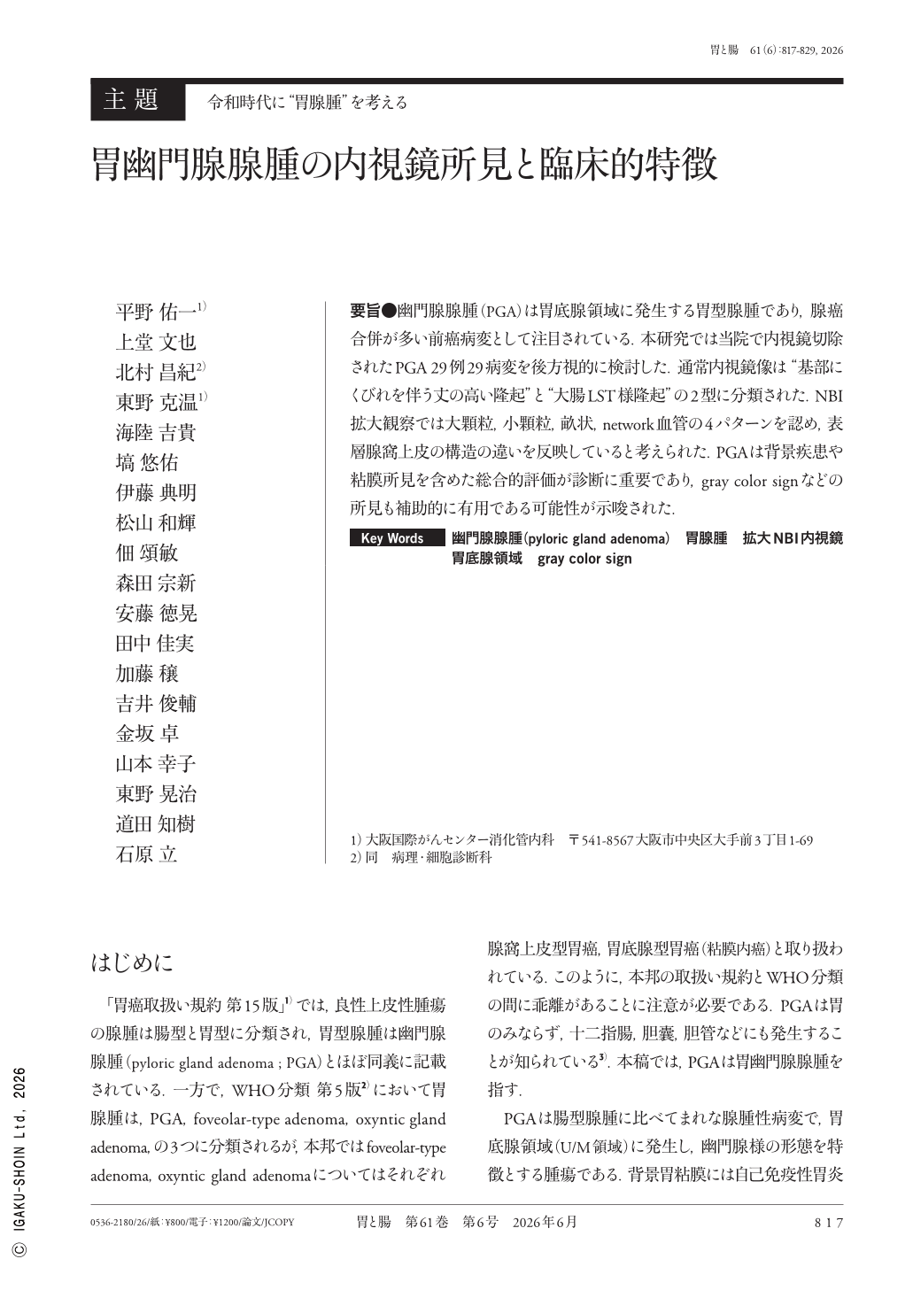
要旨●幽門腺腺腫(PGA)は胃底腺領域に発生する胃型腺腫であり,腺癌合併が多い前癌病変として注目されている.本研究では当院で内視鏡切除されたPGA 29例29病変を後方視的に検討した.通常内視鏡像は“基部にくびれを伴う丈の高い隆起”と“大腸LST様隆起”の2型に分類された.NBI拡大観察では大顆粒,小顆粒,畝状,network血管の4パターンを認め,表層腺窩上皮の構造の違いを反映していると考えられた.PGAは背景疾患や粘膜所見を含めた総合的評価が診断に重要であり,gray color signなどの所見も補助的に有用である可能性が示唆された.
Pyloric gland adenoma(PGA), a rare gastric-type adenoma that arises in the fundic gland region, is recognized as a precancerous lesion due to its frequent association with adenocarcinoma. In this study, we retrospectively analyzed 29 PGA lesions resected endoscopically at our institution. There were two categories of conventional endoscopic findings:a polypoid lesion with a slightly constricted base and a laterally spreading tumor-like morphology. Magnifying narrow-band imaging identified four patterns—large granular, small granular, ridge-like, and network vascular—reflecting differences in the surface foveolar structure. Endoscopic diagnosis of PGA requires a comprehensive evaluation that includes background diseases(such as familial adenomatous polyposis or gastric adenocarcinoma and proximal polyposis of the stomach), background mucosa(non-atrophic or atrophic), and lesion morphology. In addition, findings such as the gray color sign may serve as useful adjunctive features.
Copyright © 2026, Igaku-Shoin Ltd. All rights reserved.