Japanese
English
- 有料閲覧
- Abstract 文献概要
- 1ページ目 Look Inside
- 参考文献 Reference
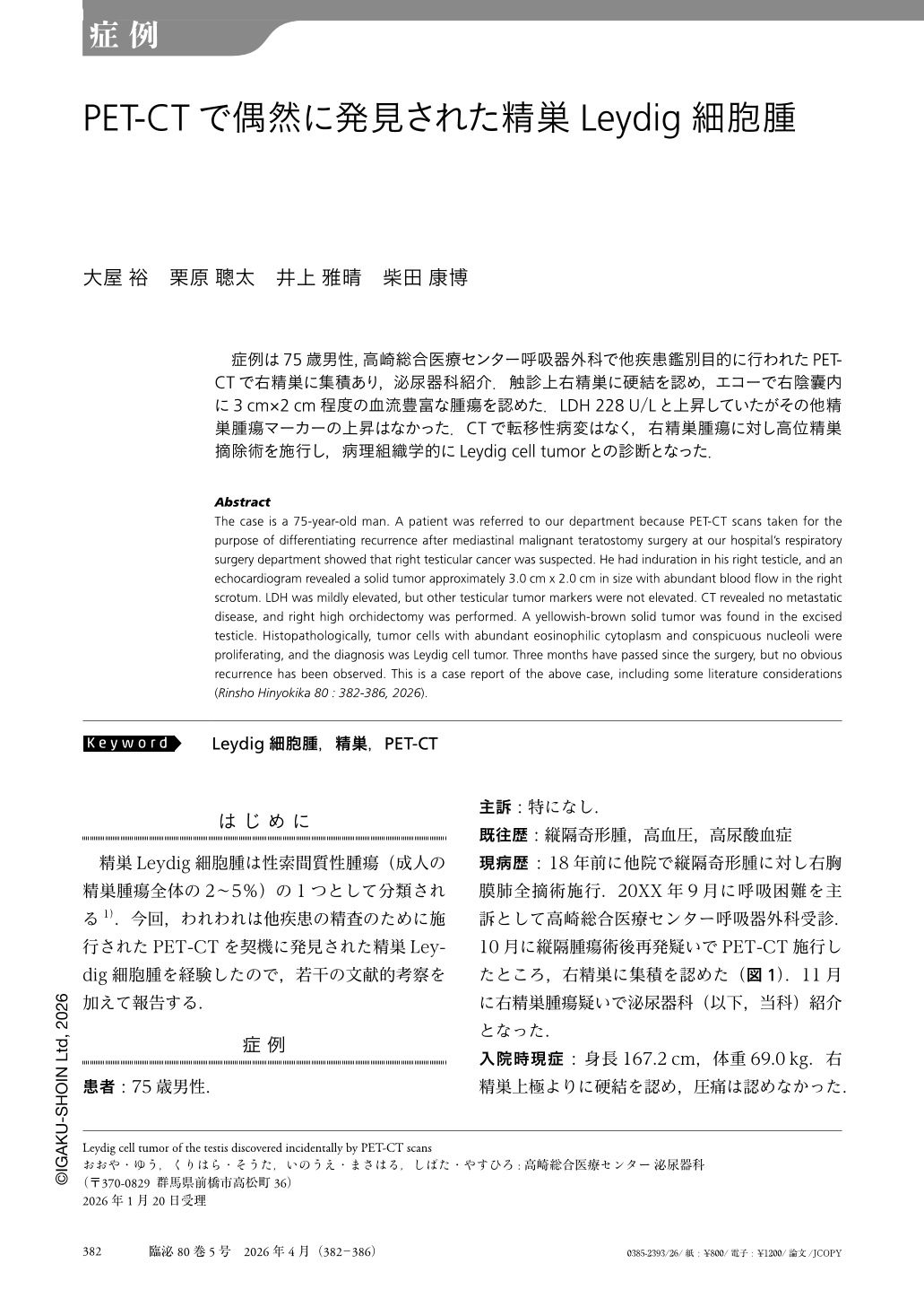
症例は75歳男性,高崎総合医療センター呼吸器外科で他疾患鑑別目的に行われたPET-CTで右精巣に集積あり,泌尿器科紹介.触診上右精巣に硬結を認め,エコーで右陰囊内に3cm×2cm程度の血流豊富な腫瘍を認めた.LDH 228U/Lと上昇していたがその他精巣腫瘍マーカーの上昇はなかった.CTで転移性病変はなく,右精巣腫瘍に対し高位精巣摘除術を施行し,病理組織学的にLeydig cell tumorとの診断となった.
Abstract
The case is a 75-year-old man. A patient was referred to our department because PET-CT scans taken for the purpose of differentiating recurrence after mediastinal malignant teratostomy surgery at our hospital's respiratory surgery department showed that right testicular cancer was suspected. He had induration in his right testicle, and an echocardiogram revealed a solid tumor approximately 3.0cm x 2.0cm in size with abundant blood flow in the right scrotum. LDH was mildly elevated, but other testicular tumor markers were not elevated. CT revealed no metastatic disease, and right high orchidectomy was performed. A yellowish-brown solid tumor was found in the excised testicle. Histopathologically, tumor cells with abundant eosinophilic cytoplasm and conspicuous nucleoli were proliferating, and the diagnosis was Leydig cell tumor. Three months have passed since the surgery, but no obvious recurrence has been observed. This is a case report of the above case, including some literature considerations (Rinsho Hinyokika 80 : 382-386, 2026).
Copyright © 2026, Igaku-Shoin Ltd. All rights reserved.