Japanese
English
- 有料閲覧
PPV購入案内
1,650円 (1,500円+税10%) こちらは、727ページから732ページの文献です。購入には医書.jp本会員登録が必要です。
会員登録完了後に改めて上記「購入サイトへ」を選択してください。
または「購入サイトへ」選択後に「購入ログイン」、「新規会員登録」の順に進んでください。
会員登録について詳しくはをご確認ください。
- Abstract 文献概要
- 1ページ目 Look Inside
- 参考文献 Reference
- サイト内被引用 Cited by
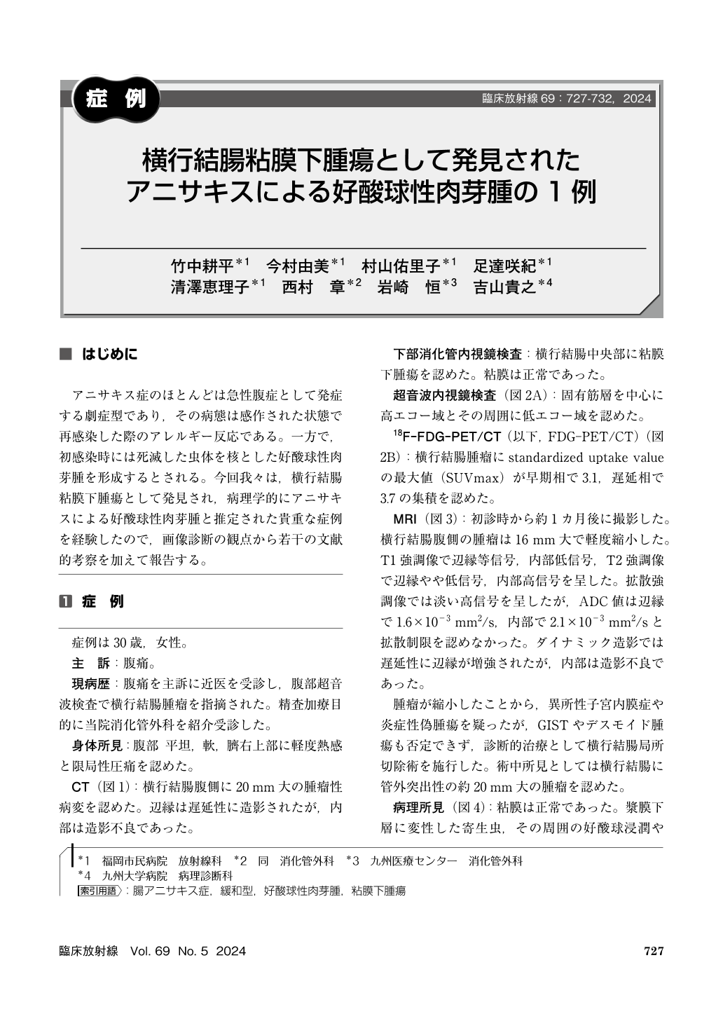
アニサキス症のほとんどは急性腹症として発症する劇症型であり,その病態は感作された状態で再感染した際のアレルギー反応である。一方で,初感染時には死滅した虫体を核とした好酸球性肉芽腫を形成するとされる。今回我々は,横行結腸粘膜下腫瘍として発見され,病理学的にアニサキスによる好酸球性肉芽腫と推定された貴重な症例を経験したので,画像診断の観点から若干の文献的考察を加えて報告する。
A 30–year–old woman with abdominal pain was referred to our hospital after abdominal ultrasound revealed a transverse colon mass. The contrast–enhanced CT and MRI scans showed the mass, whose rim is enhanced but whose inside is not. 18F–FDG–PET/CT showed weak 18F–FDG accumulation. Histopathological examination revealed an eosinophilic granuloma around a degenerative parasite consistent with the characteristics of anisakiasis. Chronic anisakiasis typically forms a submucosal tumor–like lesion and shows pathologically an eosinophilic granuloma around an anisakis larva. Because it is rare and its imaging findings are not established, it is occasionally misdiagnosed and removed by endoscopic or surgical resection. We report the radiological features of the eosinophilic granuloma related to anisakiasis and review the literature on it.
Copyright © 2024, KANEHARA SHUPPAN Co.LTD. All rights reserved.