Japanese
English
- 有料閲覧
PPV購入案内
2,200円 (2,000円+税10%) こちらは、634ページから637ページの文献です。購入には医書.jp本会員登録が必要です。
会員登録完了後に改めて上記「購入サイトへ」を選択してください。
または「購入サイトへ」選択後に「購入ログイン」、「新規会員登録」の順に進んでください。
会員登録について詳しくはをご確認ください。
- Abstract 文献概要
- 1ページ目 Look Inside
- 参考文献 Reference
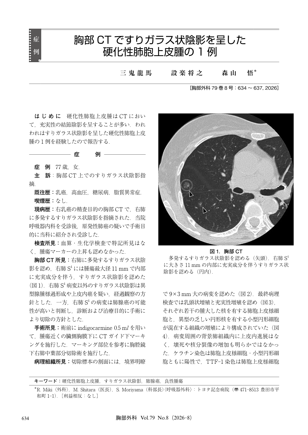
はじめに 硬化性肺胞上皮腫はCTにおいて,充実性の結節陰影を呈することが多い.われわれはすりガラス状陰影を呈した硬化性肺胞上皮腫の1例を経験したので報告する.
A 77-year-old woman indicated multiple ground-glass opacities (GGOs) in the right lung on chest computed tomography (CT). A 11 mm GGO in S4 of the right lung showed a part solid component. Primary lung cancer was suspected, and surgical resection was planned. After CT-guided marking, video-assisted thoracoscopic partial resection of the right middle lobe was undergone. The postoperative course was uneventful, and the patient was discharged on postoperative day three. Histopathological examination showed proliferation of alveolar epithelial cells with mildly enlarged nuclei and small round cells with minimal atypia, leading to a diagnosis of pulmonary sclerosing pneumocytoma. This case highlights that pulmonary sclerosing pneumocytoma can present as an atypical GGO on chest CT.
© Nankodo Co., Ltd., 2026