Japanese
English
- 有料閲覧
PPV購入案内
1,650円 (1,500円+税10%) こちらは、573ページから577ページの文献です。購入には医書.jp本会員登録が必要です。
会員登録完了後に改めて上記「購入サイトへ」を選択してください。
または「購入サイトへ」選択後に「購入ログイン」、「新規会員登録」の順に進んでください。
会員登録について詳しくはをご確認ください。
- Abstract 文献概要
- 1ページ目 Look Inside
- 参考文献 Reference
- サイト内被引用 Cited by
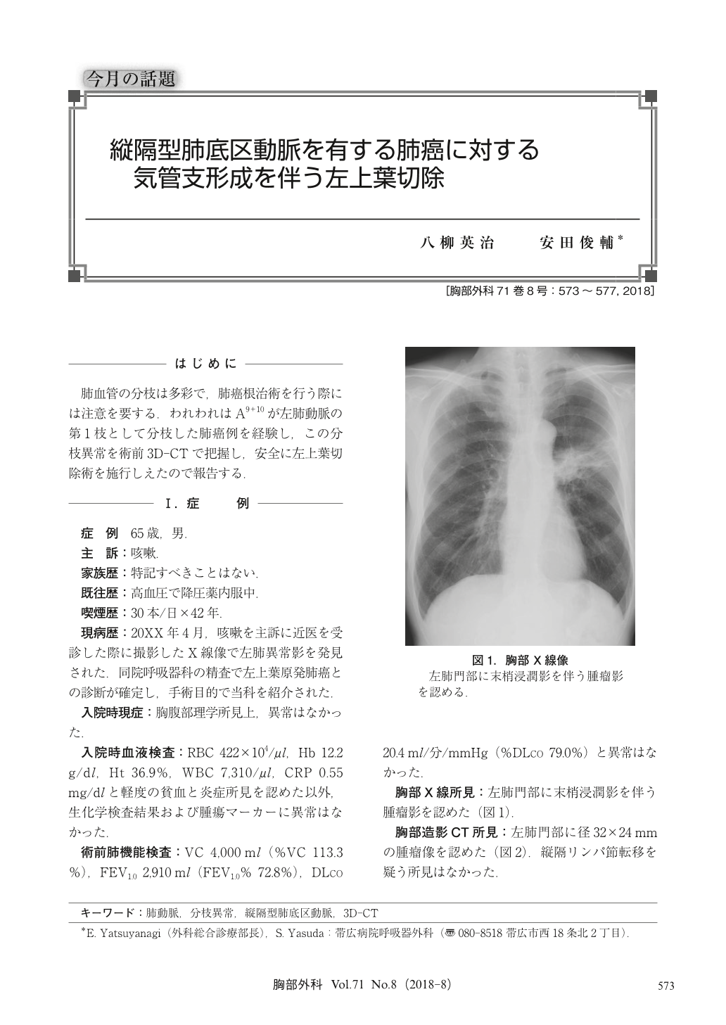
肺血管の分枝は多彩で,肺癌根治術を行う際には注意を要する.われわれはA9+10が左肺動脈の第1枝として分枝した肺癌例を経験し,この分枝異常を術前3D-CTで把握し,安全に左上葉切除術を施行しえたので報告する.
A 65-year-old man with left lung squamous cell carcinoma was admitted to our hospital for operation. His tumor was located left upper lobe and invaded to the orifice of left upper bronchus. We diagnosed the tumor as cT2aN0M0 and intended to perform radical operation. Preoperative three-dimensional computed tomography (3D-CT) revealed that A9+10 which descended along lower bronchus was arisen from the left main pulmonary artery as a 1st branch of it. A wedge bronchoplastic left upper lobectomy was safely done with preserving the branch. Although this abnormal branching of left pulmonary artery is very rare, we should pay more attention this type of anomaly because unintended injury of the branch causes massive bleeding or ischemia of left lower lobe. Preoperative 3D-CT is useful for detecting the anomaly of pulmonary vessels.
© Nankodo Co., Ltd., 2018