Japanese
English
- 有料閲覧
PPV購入案内
1,320円 (1,200円+税10%) こちらは、359ページから365ページの文献です。購入には医書.jp本会員登録が必要です。
会員登録完了後に改めて上記「購入サイトへ」を選択してください。
または「購入サイトへ」選択後に「購入ログイン」、「新規会員登録」の順に進んでください。
会員登録について詳しくはをご確認ください。
- Abstract 文献概要
- 1ページ目 Look Inside
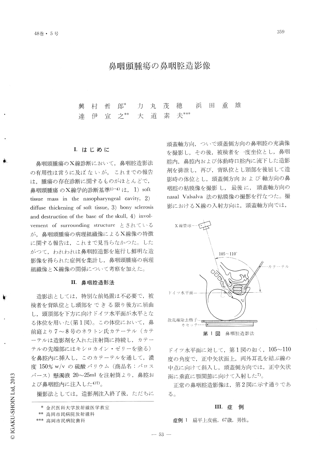
I.はじめに
鼻咽頭腫瘍のX線診断において,鼻咽腔造影法の有用性は言うに及ばないが,これまでの報告は,腫瘍の存在診断に関するものがほとんどで,鼻咽頭腫瘍のX線学的診断基準1)〜4)は,1)soft tissue mass in the nasopharyngeal cavity,2)diffuse thickening of soft tissue,3)bony sclerosis and destruction of the base of the skull,4)involvement of surrounding structureとされているが,鼻咽頭腫瘍の病理組織像によるX線像の特徴に関する報告は,これまで見当らなかつた。したがつて,われわれは鼻咽腔造影を施行し鮮明な造影像を得られた症例を集計し,鼻咽頭腫瘍の病理組織像とX線像の関係について考察を加えた。
A study on the nasopharyngeal tumors, consisting of 6 cases of epidermoid carcinomas, 7 cases ofreticulum cell sarcomas and others, a total of 17 cases, by means of contrast nasopharyngography by using barium sulfate, is reported.
The nasopharyngograms of the carcinomas showed a diffuse thickening of the soft tissue with underfianable margins and irregular surfacecontour, and also accompanied by evidence of bony destructions in many cases.
On the other hand, in cases of sarcomas the tumor mass was sharply defined with irregular surface, but there was no evidence of bony involvement.
Copyright © 1976, Igaku-Shoin Ltd. All rights reserved.