Japanese
English
- 有料閲覧
PPV購入案内
1,320円 (1,200円+税10%) こちらは、249ページから253ページの文献です。購入には医書.jp本会員登録が必要です。
会員登録完了後に改めて上記「購入サイトへ」を選択してください。
または「購入サイトへ」選択後に「購入ログイン」、「新規会員登録」の順に進んでください。
会員登録について詳しくはをご確認ください。
- Abstract 文献概要
- 1ページ目 Look Inside
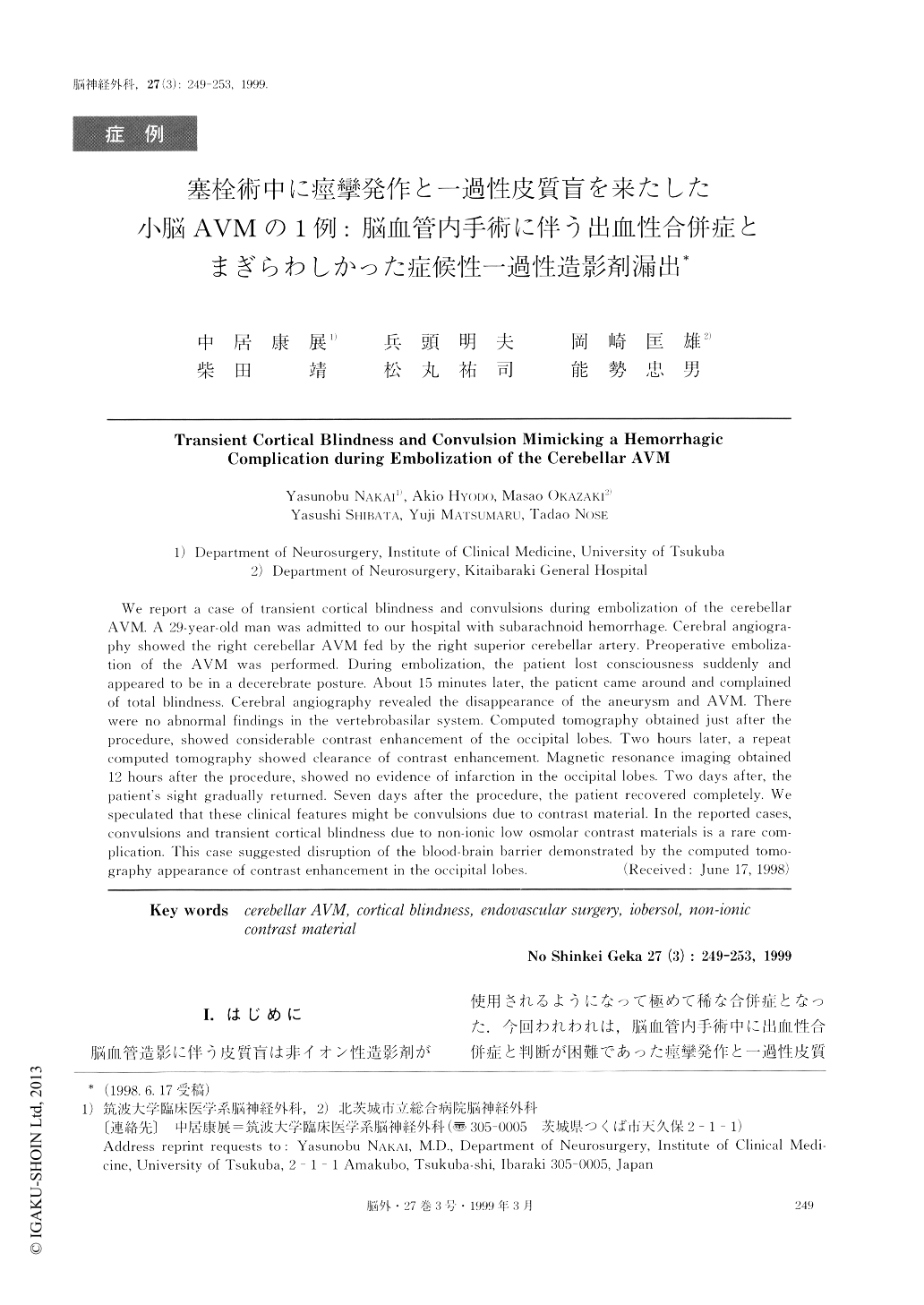
I.はじめに
脳血管造影に伴う皮質盲は非イオン性造影剤が使用されるようになって極めて稀な合併症となった.今回われわれは,脳血管内手術中に出血性合併症と判断が困難であった痙攣発作と一過性皮質盲を呈した小脳AVMの1例を経験した.本例における特徴的な画像所見を含め,若干の文献的考察を加えて報告する.
We report a case of transient cortical blindness and convulsions during embolization of the cerebellarAVM. A 29-year-old man was admitted to our hospital with subarachnoid hemorrhage. Cerebral angiogra-phy showed the right cerebellar AVM fed by the right superior cerebellar artery. Preoperative emboliza-tion of the AVM was performed. During embolization, the patient lost consciousness suddenly andappeared to be in a decerebrate posture. About 15 minutes later, the patient came around and complainedof total blindness. Cerebral angiography revealed the disappearance of the aneurysm and AVM. Therewere no abnormal findings in the vertebrobasilar system. Computed tomography obtained just after theprocedure, showed considerable contrast enhancement of the occipital lobes. Two hours later, a repeatcomputed tomography showed clearance of contrast enhancement. Magnetic resonance imaging obtained12 hours after the procedure, showed no evidence of infarction in the occipital lobes. Two days after, thepatient's sight gradually returned. Seven days after the procedure, the patient recovered completely. Wespeculated that these clinical features might be convulsions due to contrast material. In the reported cases,convulsions and transient cortical blindness clue to non-ionic low osmolar contrast materials is a rare com-plication. This case suggested disruption of the blood-brain barrier demonstrated by the computed tomo-graphy appearance of contrast enhancement in the occipital lobes.
Copyright © 1999, Igaku-Shoin Ltd. All rights reserved.