Japanese
English
- 有料閲覧
PPV購入案内
1,320円 (1,200円+税10%) こちらは、843ページから848ページの文献です。購入には医書.jp本会員登録が必要です。
会員登録完了後に改めて上記「購入サイトへ」を選択してください。
または「購入サイトへ」選択後に「購入ログイン」、「新規会員登録」の順に進んでください。
会員登録について詳しくはをご確認ください。
- Abstract 文献概要
- 1ページ目 Look Inside
- 参考文献 Reference
Ⅰ.はじめに
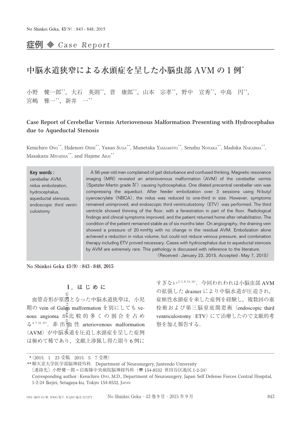
血管奇形が原因となった中脳水道狭窄は,小児期のvein of Galen malformationを別にしてもvenous angiomaが比較的多くの割合を占める4,7,21,23).非出血性arteriovenous malformation(AVM)が中脳水道を圧迫し水頭症を呈した症例は極めて稀であり,文献上渉猟し得た限り6例にすぎない2,3,9,11,14).今回われわれは小脳虫部AVMの拡張したdrainerにより中脳水道が圧迫され,症候性水頭症を来した症例を経験し,複数回の塞栓術および第三脳室底開窓術(endoscopic third ventriculostomy:ETV)にて治療したので文献的考察を加え報告する.
A 56-year-old man complained of gait disturbance and confused thinking. Magnetic resonance imaging(MRI)revealed an arteriovenous malformation(AVM)of the cerebellar vermis(Spetzler-Martin grade Ⅳ)causing hydrocephalus. One dilated precentral cerebellar vein was compressing the aqueduct. After feeder embolization over 3 sessions using N-butyl cyanoacrylate(NBCA), the nidus was reduced to one-third in size. However, symptoms remained unimproved, and endoscopic third ventriculostomy(ETV)was performed. The third ventricle showed thinning of the floor, with a fenestration in part of the floor. Radiological findings and clinical symptoms improved, and the patient returned home after rehabilitation. The condition of the patient remained stable as of six months later. On angiography, the draining vein showed a pressure of 20mmHg with no change in the residual AVM. Embolization alone achieved a reduction in nidus volume, but could not reduce venous pressure, and combination therapy including ETV proved necessary. Cases with hydrocephalus due to aqueductal stenosis by AVM are extremely rare. This pathology is discussed with reference to the literature.
Copyright © 2015, Igaku-Shoin Ltd. All rights reserved.