Japanese
English
- 有料閲覧
PPV購入案内
1,320円 (1,200円+税10%) こちらは、265ページから269ページの文献です。購入には医書.jp本会員登録が必要です。
会員登録完了後に改めて上記「購入サイトへ」を選択してください。
または「購入サイトへ」選択後に「購入ログイン」、「新規会員登録」の順に進んでください。
会員登録について詳しくはをご確認ください。
- Abstract 文献概要
- 1ページ目 Look Inside
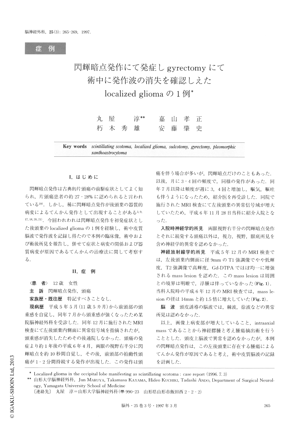
I.はじめに
閃輝暗点発作は古典的片頭痛の前駆症状としてよく知られ,片頭痛患者の約27-28%に認められると言われている19).しかし,稀に閃輝暗点発作が後頭葉の器質的病変によるてんかん発作として出現することがある3,9,17,18,20,21).今回われわれは閃輝暗点発作を初発症状とした後頭葉のlocalized gliomaの1例を経験し,術中皮質脳波で発作波を記録し得たので本例の臨床像,術中および術後所見を報告し,併せて症状と病変の関係および器質病変が原因であるてんかんの治療法に関して考察する.
An 11-year-old female presented with headache in May 1993. Magnetic resonance (MR) imaging disclosed a small lesion (9mm in diameter) in the left occipital lobe. No treatment was performed because the lesion was small. She subsequently developed frequent epi-sodes of scintillating scotoma in the right visual field eleven months later. MR imaging eleven months after the first MR imaging showed the lesion had enlarged to 14mm in diameter. Preoperative surface elec-troencephalography (EEG) detected no spike waves. The diagnosis was localized glioma. The mass was totally removed by gyrectomy in December 1994. In-traoperative cortical EEG demonstrated spike waves which disappeared after tumor removal. The histologic-al diagnosis was pleomorphic xanthoastrocytoma. No postoperative neurological deficit was recognized, and scintillating scotoma and headache disappeared. Postop-erative stereotactic radiosurgery was performed. The scintillating scotoma was caused by the tumor, because the spike wave and phase reversal were detected by the intraoperative cortical EEG. Intraoperative EEG is use-ful for the diagnosis of epilepsy caused by tumor. Sul-cotomy and gyrectomy are the optimal surgical treat-ments for epilepsy caused by a localized glioma.
Copyright © 1997, Igaku-Shoin Ltd. All rights reserved.