Japanese
English
- 有料閲覧
PPV購入案内
1,320円 (1,200円+税10%) こちらは、1135ページから1138ページの文献です。購入には医書.jp本会員登録が必要です。
会員登録完了後に改めて上記「購入サイトへ」を選択してください。
または「購入サイトへ」選択後に「購入ログイン」、「新規会員登録」の順に進んでください。
会員登録について詳しくはをご確認ください。
- Abstract 文献概要
- 1ページ目 Look Inside
I.はじめに
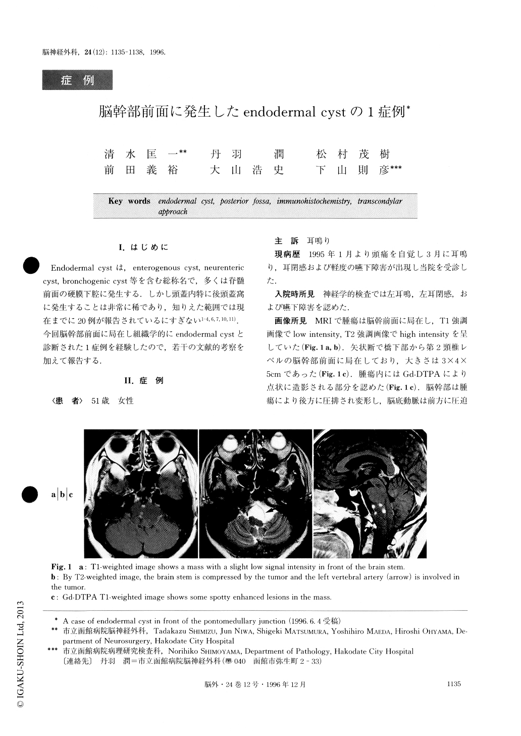
Endodermal cystは,enterogenous cyst,neurenteric cyst,bronchogenic cyst等を含む総称名で,多くは脊髄前面の硬膜下腔に発生する.しかし頭蓋内特に後頭蓋窩に発生することは非常に稀であり,知りえた範囲では現在までに20例が報告されているにすぎない1-4,6,7,10,11).今回脳幹部前面に局在し組織学的にendodermal cystと診断された1症例を経験したので,若干の文献的考察を加えて報告する.
We reported a rare case of an endodermal cyst on the posterior fossa. The patient was a 51-year-old woman with tinnitus, ear blockage and a mild swallow-ing disturbance. MR images revealed a cystic mass 5 cm in diameter in front of the pontomedullary junction. Gadolinium-enhanced images showed some spotty le-sions in the cystic mass. The brain stem was compres-sed strongly by the mass. The left vertebral artery was involved in the mass. Total removal of the tumor was performed via the transcondylar approach. During the operation the lower cranial nerves, the bilateral verte-bral artery and the vertebral union were recognized. The cystic mass consisted of a yellow colored thin wall and a watery fluid which contained some small hard lesions. Histopathological examination of the cyst wall revealed a single layer of ciliated columnar epithelium, and that of the small lesions showed histiocytes and granulation. Immunohistochemically, the cyst wall was stained by an epithelial membrane antigen and a carcinoembryonic antigen. From these histopathological findings, the final diagnosis of the cystic lesion was an endodermal cyst. Twenty such cases have been reported to date since Afshar reported a case of an endodermal cyst on the posterior fossa in 1981. There are many types of cystic lesions in the intracranial region, and immunohistoche-mical studies are necessary for diagnosis of these in-tracranial cysts.
Copyright © 1996, Igaku-Shoin Ltd. All rights reserved.