Japanese
English
- 有料閲覧
- Abstract 文献概要
- 1ページ目 Look Inside
- 参考文献 Reference
Ⅰ.はじめに
内胚葉囊胞(endodermal cyst)は,呼吸器や腸管の上皮に類似した細胞からなる囊胞であり,胎生第3週頃の外胚葉と内胚葉の分離不全が原因とされ,全中枢神経系腫瘍の0.01%と非常に稀な疾患である1,6,7).今回われわれは,進行性の右上下肢不全麻痺で発症した頸髄endodermal cystに対し,囊胞壁の亜全摘出術を行い良好な経過を得た1例を経験したので,文献的考察を加えて報告する.
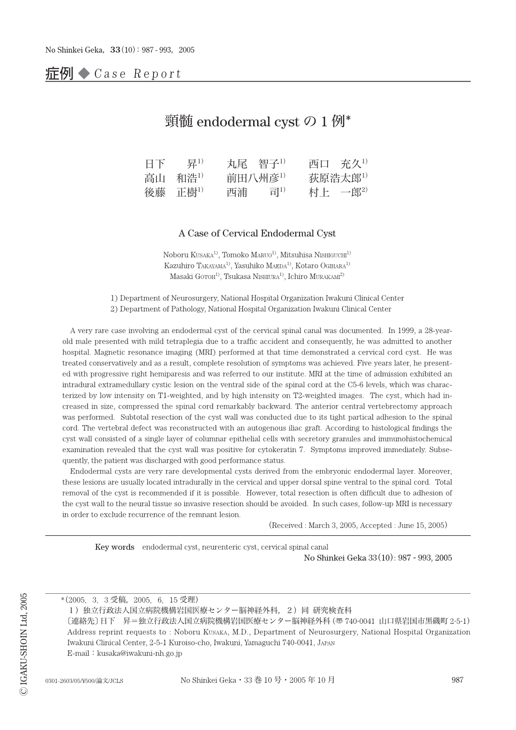
A very rare case involving an endodermal cyst of the cervical spinal canal was documented. In 1999,a 28-year-old male presented with mild tetraplegia due to a traffic accident and consequently,he was admitted to another hospital. Magnetic resonance imaging (MRI) performed at that time demonstrated a cervical cord cyst. He was treated conservatively and as a result,complete resolution of symptoms was achieved. Five years later,he presented with progressive right hemiparesis and was referred to our institute. MRI at the time of admission exhibited an intradural extramedullary cystic lesion on the ventral side of the spinal cord at the C5-6 levels,which was characterized by low intensity on T1-weighted,and by high intensity on T2-weighted images. The cyst,which had increased in size,compressed the spinal cord remarkably backward. The anterior central vertebrectomy approach was performed. Subtotal resection of the cyst wall was conducted due to its tight partical adhesion to the spinal cord. The vertebral defect was reconstructed with an autogenous iliac graft. According to histological findings the cyst wall consisted of a single layer of columnar epithelial cells with secretory granules and immunohistochemical examination revealed that the cyst wall was positive for cytokeratin 7. Symptoms improved immediately. Subsequently,the patient was discharged with good performance status.
Endodermal cysts are very rare developmental cysts derived from the embryonic endodermal layer. Moreover,these lesions are usually located intradurally in the cervical and upper dorsal spine ventral to the spinal cord. Total removal of the cyst is recommended if it is possible. However,total resection is often difficult due to adhesion of the cyst wall to the neural tissue so invasive resection should be avoided. In such cases,follow-up MRI is necessary in order to exclude recurrence of the remnant lesion.

Copyright © 2005, Igaku-Shoin Ltd. All rights reserved.


